What this guide is for
This guide is about cell division: how one cell becomes two cells. It covers the two kinds of cell division that happen in plants and animals, called mitosis and meiosis, and the simpler kind that bacteria use, called binary fission.
Read the parts in order. Part 2 explains chromosomes, and everything after it depends on that part. The drawings of dividing cells show only 2 pairs of chromosomes instead of the 23 pairs in a human cell, so that you can count every chromosome and follow where it goes.
At the end there are questions that check themselves, and a list of the terms used in the guide.
Goes with: Look Again Quiz 13: Cell Division · Life Science: Start Here, Part 6
In this guide:
Why cells divide
Cell division is how living things grow, repair damage, and reproduce. When a cell divides, one cell becomes two cells.
There are two main types of cell division you need to know for the GED:
- Mitosis makes identical body cells, for growth and repair. A body cell is any cell in the body that is not a sex cell: skin cells, muscle cells, and bone cells are all body cells.
- Meiosis makes sex cells (sperm and eggs), for reproduction.
Bacteria use a third, simpler kind of division, called binary fission. It is explained in Part 6.
All cells come from cells that already existed, through cell division. New cells do not form from nonliving material. The German doctor Rudolf Virchow made this idea famous in 1855, and it is one of the basic principles of biology.
Understanding chromosomes first
Before we look at cell division, you need to understand what chromosomes are, because chromosomes are what a dividing cell has to divide fairly between the new cells.
Chromosomes are tightly coiled structures made of DNA wound around proteins. DNA carries the genetic information: the instructions for making you.
- Human body cells have 46 chromosomes (23 pairs).
- These come in matching pairs. In each pair, one chromosome came from your mother and one came from your father. The two chromosomes of a pair are called homologous chromosomes ("homologous" means matching): they carry genes for the same traits, in the same order.
- We call this the diploid number (2n), because the chromosomes come in pairs. "Di" means two: a body cell has two full sets of 23.
- Sex cells (sperm and egg) have only 23 chromosomes, half the number in a body cell. A sex cell gets one chromosome from each pair, so it has one full set of 23.
- This number, one set, is called the haploid number (n).
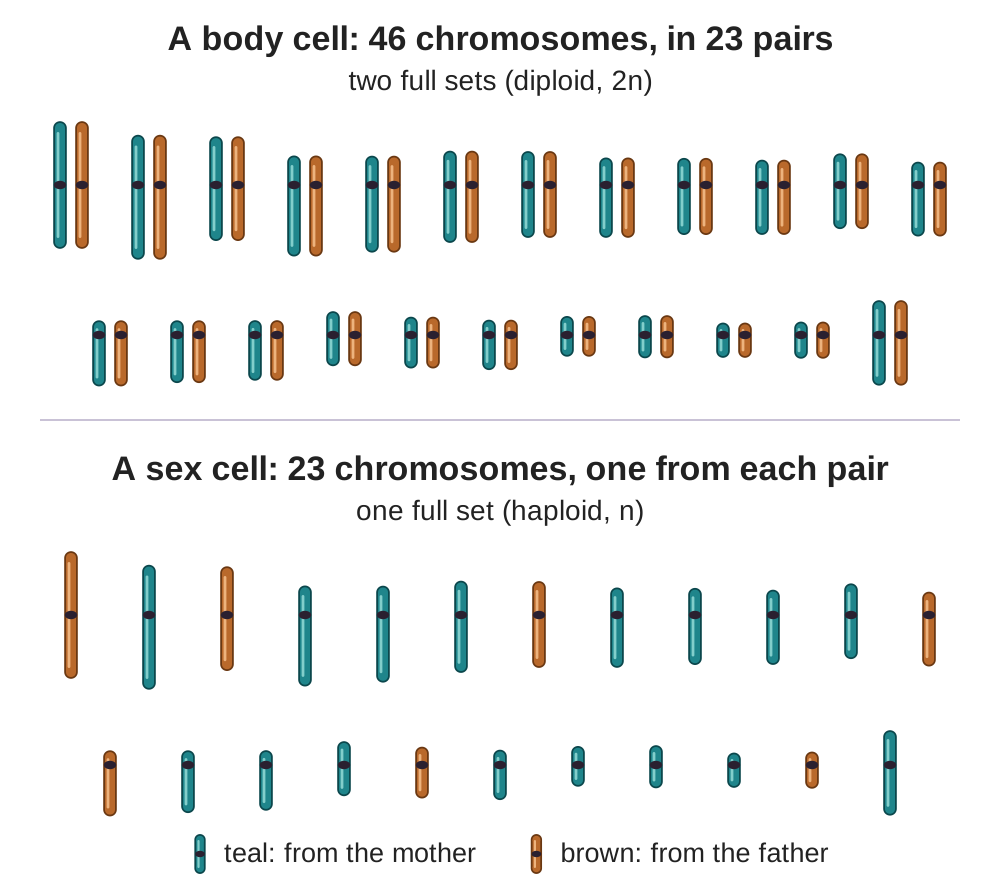
The two numbers fit together. When a sperm with 23 chromosomes joins an egg with 23 chromosomes (this joining is called fertilization), the fertilized egg has 23 + 23 = 46 chromosomes: the right number for a body cell. Every body cell of the new person comes from that one cell, by mitosis.
Mitosis: making identical body cells
- Purpose: growth, repair, and replacement of body cells.
- Result: 2 identical daughter cells, each with 46 chromosomes (diploid). "Daughter cells" is simply the name biologists use for the new cells made by a division.
- Where: in body cells, which means all cells except the sex cells. Not every kind of body cell keeps dividing: most nerve cells in an adult, for example, rarely divide.
Before mitosis: each chromosome is copied
A cell spends most of its life not dividing. During this time, called interphase, the cell grows and does its work. Before it divides, it makes a copy of all of its DNA.
After copying, each chromosome is made of two identical strands joined at one point. Each strand is called a sister chromatid, and the point where the two are joined is called the centromere. That is why chromosomes in pictures of dividing cells look like an X: each X is two copies held together. (In this guide’s drawings, the two copies are drawn side by side.) Mitosis pulls the two copies apart, so that each new cell gets one.
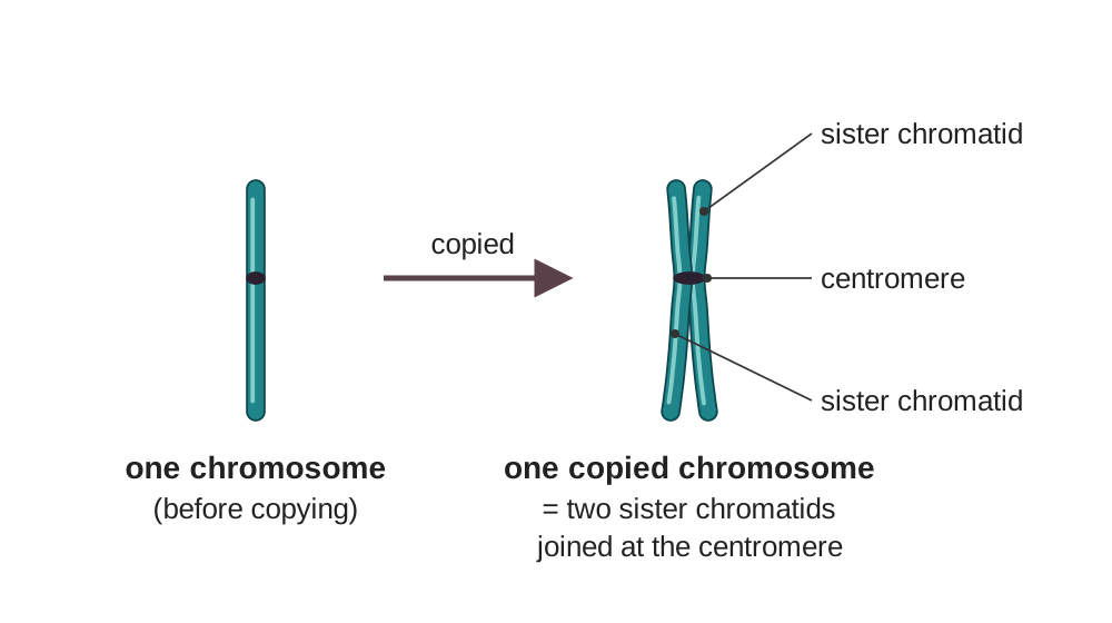
A real human cell has 46 chromosomes, which is too many to count in a drawing. The drawings show a simplified cell with only 4 chromosomes: 2 pairs. The long pair and the short pair are different chromosomes. In each pair, the teal chromosome came from the mother and the brown one came from the father. All the drawings use the same sizes, so a long chromosome is the same length in every picture.
The four phases of mitosis
Mitosis is described in four stages, called phases. They happen in this order: prophase, metaphase, anaphase, telophase. In a real cell, each phase flows into the next without a pause; the names mark points along the way.
1. Prophase
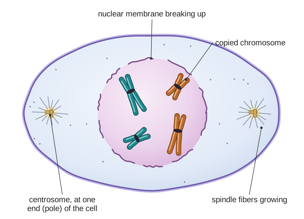
The chromatin (the loose, thread-like form the DNA takes when the cell is not dividing) coils up tightly into chromosomes you can see under a microscope, each shaped like an X. The nuclear membrane (the thin envelope around the nucleus) breaks down. Spindle fibers begin to form. Spindle fibers are thin threads made of protein that stretch across the cell; they are what will move the chromosomes. "Pro" means before: prophase is the first phase.
2. Metaphase
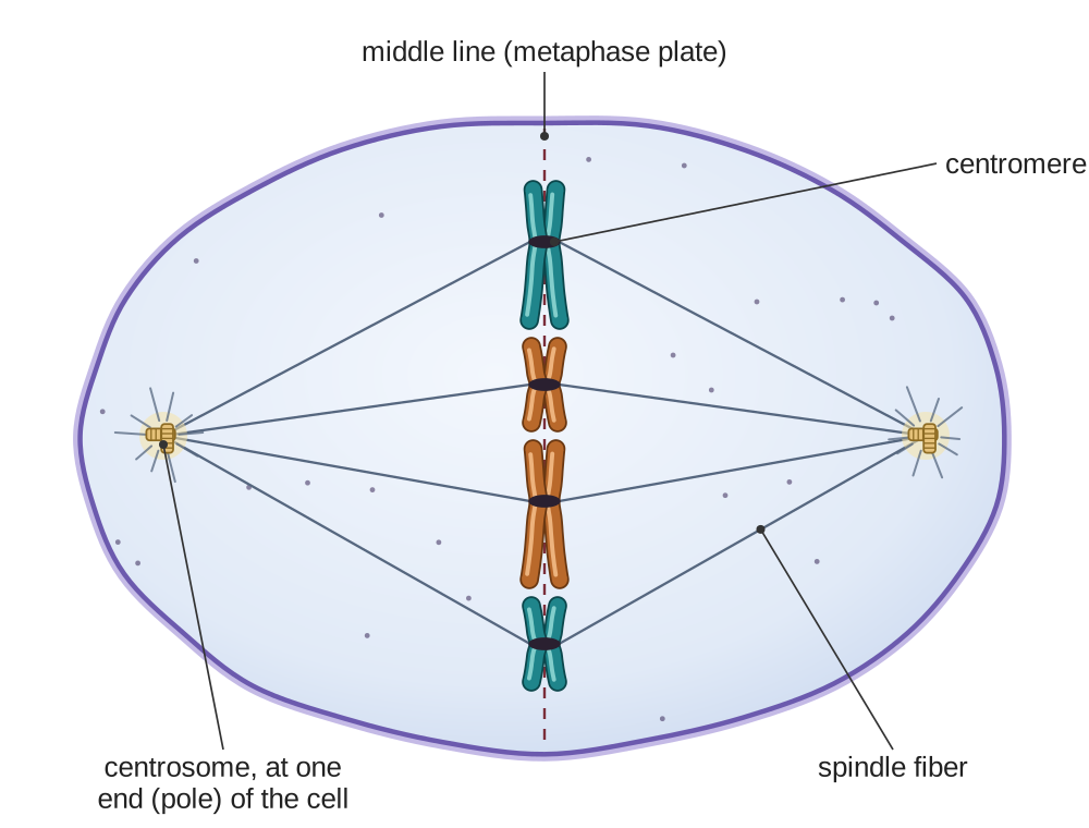
Chromosomes line up single file along the middle (the equator) of the cell. Spindle fibers attach to the centromere of each chromosome, the point where the two sister chromatids are joined, with fibers coming from both ends of the cell. The middle line is sometimes called the metaphase plate. To remember it: M for Middle.
3. Anaphase
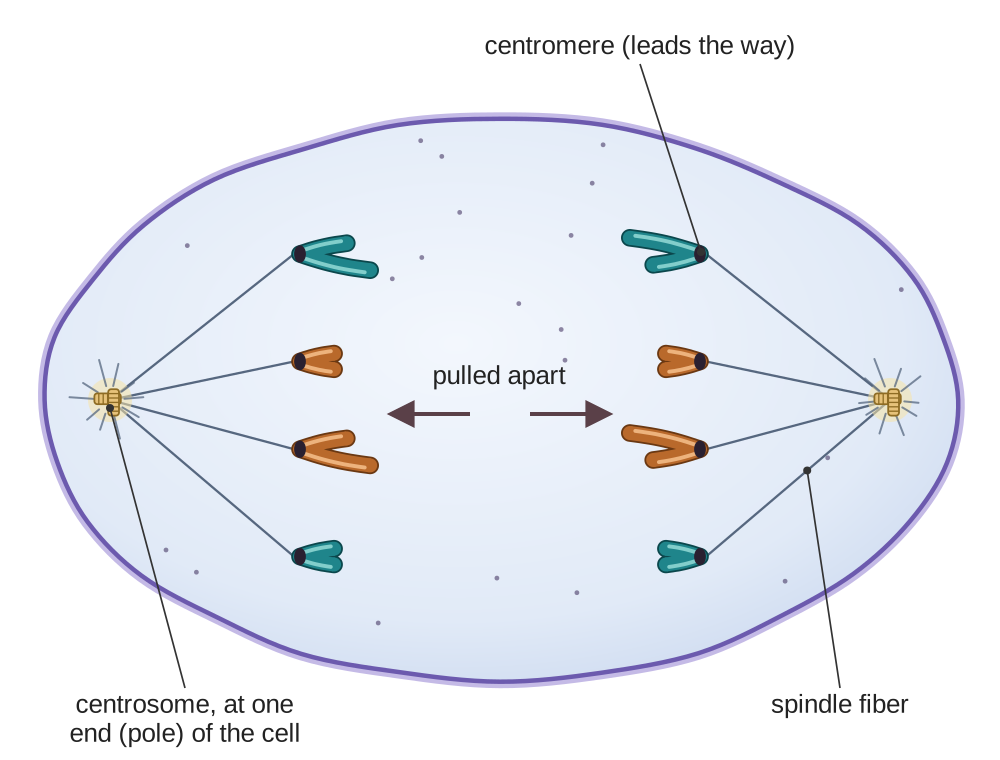
Sister chromatids (the two halves of each X) are pulled apart to opposite ends of the cell by the spindle fibers. Once they are separated, each chromatid counts as a chromosome of its own. Each end of the cell now has one complete set: in a human cell, 46 chromosomes at each end. To remember it: A for Away from each other.
4. Telophase and cytokinesis
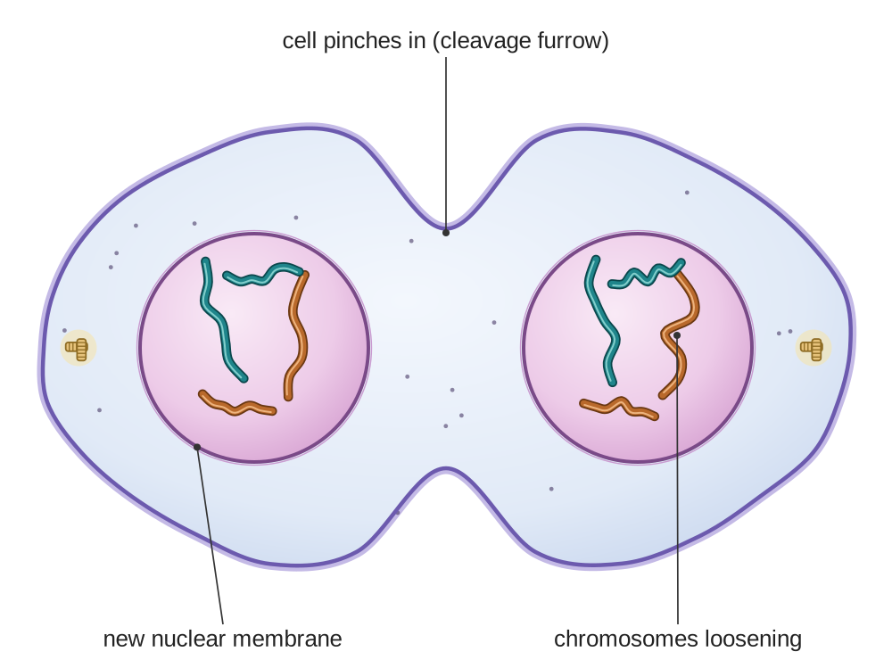
Nuclear membranes form around each set of chromosomes. The chromosomes uncoil and become loose chromatin again. The cell pinches in at the middle to create two separate cells. The pinched-in groove is called the cleavage furrow. This splitting of the cell itself is called cytokinesis ("cyto" means cell); it usually begins while telophase is still going on. "Telo" means end: telophase is the last phase, and two new cells are forming.
Plant cells have a stiff cell wall and cannot pinch in. Instead, a new wall, called the cell plate, grows across the middle of the cell and divides it in two.
A memory aid for the order of the phases
Once you know what happens in each phase, a sentence can help you keep the phases in order. The first letters of "Please Make A Telephone" (or "Please Meet At Three") are P, M, A, T: Prophase, Metaphase, Anaphase, Telophase. This is only a memory aid. The words of the sentence have nothing to do with what happens in the cell.
Think it through. A skin cell divides by mitosis, and each of its two daughter cells has all 46 chromosomes. The parent cell had only 46. How can each new cell end up with the full 46?
Show a model answer
Before mitosis, during interphase, the cell copies all of its DNA, so each of the 46 chromosomes becomes two sister chromatids. In anaphase the sister chromatids are pulled apart, one to each end of the cell. So each new cell receives one copy of all 46 chromosomes. Nothing is split in half; everything was copied first.
Meiosis: making sex cells
- Purpose: to make sex cells, also called gametes, for reproduction.
- Result: 4 unique daughter cells, each with 23 chromosomes (haploid).
- Where: in humans, only in the ovaries and the testes.
- Special feature: meiosis creates genetic variation (differences in genes from one individual to the next). Each sex cell is different.
Why meiosis is different
As in mitosis, the cell copies its DNA once before it begins. But meiosis then has TWO divisions instead of one:
- Meiosis I separates the chromosome pairs: one chromosome of each pair goes to each new cell. This reduces the number from 46 to 23. Each of these 23 is still a copied chromosome, made of two sister chromatids.
- Meiosis II separates the sister chromatids, the way mitosis does. The result is 4 cells, each with 23 single chromosomes.
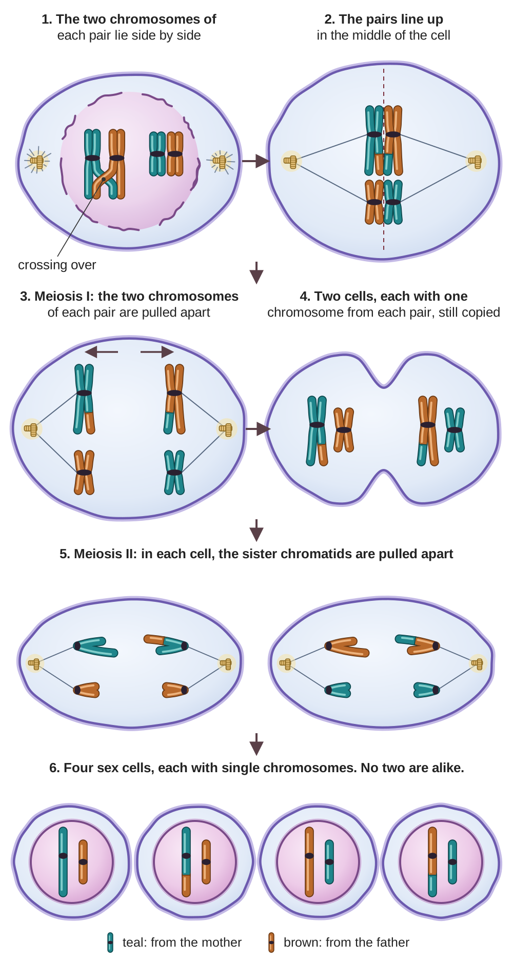
In men, all four cells become sperm. In women, the divisions are uneven: one of the cells becomes the egg and keeps most of the cytoplasm (the material inside the cell), and the other, much smaller cells break down.
Key features of meiosis
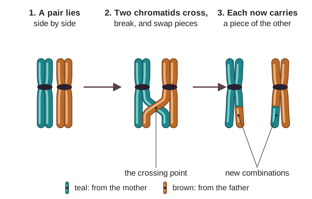
- Crossing over: during meiosis I, the two chromosomes of each pair lie side by side and swap pieces of DNA. This creates new combinations of genes on each chromosome.
- Random assortment (also called independent assortment): when the pairs separate in meiosis I, which chromosome of each pair goes into which cell is random, like a coin toss for each of the 23 pairs. This adds more variation. Counting only this coin toss, one person can make more than 8 million different combinations of chromosomes (2 multiplied by itself 23 times).
- Genetic diversity: together, these two processes make every sex cell genetically unique. That is one reason brothers and sisters with the same parents look different. (The other reason is chance: which sperm joins which egg.)
Think it through. Why do sex cells need only 23 chromosomes, and not 46?
Show a model answer
Because a new person starts when a sperm and an egg join. If each had 46, the fertilized egg would have 92, twice the right number. With 23 in each, the fertilized egg has 23 + 23 = 46, the correct number for a human body cell.
Mitosis and meiosis compared
The table puts the two kinds of division side by side.
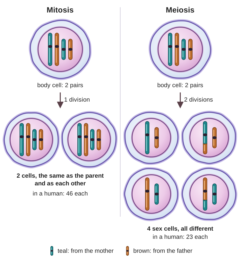
| Feature | Mitosis | Meiosis |
|---|---|---|
| Purpose | Growth, repair, replacement | Make sex cells for reproduction |
| Where it happens | Body cells | In humans, only in the ovaries and testes |
| Number of divisions | 1 division | 2 divisions |
| Cells produced | 2 daughter cells | 4 daughter cells |
| Chromosome number | 46 chromosomes (diploid) Same as parent | 23 chromosomes (haploid) Half of parent |
| Genetic identity | Identical to the parent and to each other | Genetically unique (different from the parent and from each other) |
| Crossing over? | No | Yes, in meiosis I |
Binary fission: how bacteria divide
Binary fission is much simpler than mitosis and meiosis. It happens only in prokaryotes, such as bacteria. Prokaryotes are single cells with no nucleus. You should know what binary fission is, but do not confuse it with mitosis.
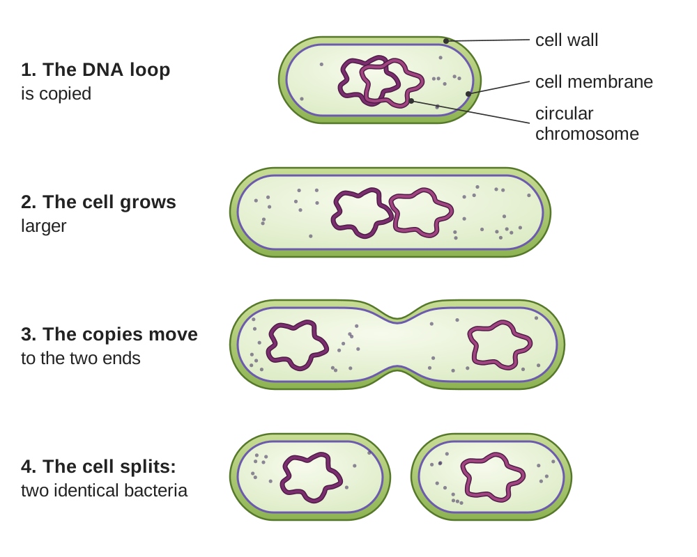
Binary fission is how bacteria reproduce. It is a simple splitting process in which one cell becomes two identical cells. ("Binary" means two; "fission" means splitting.)
How binary fission works
- The bacterial DNA (one circular chromosome, a closed loop) makes a copy of itself.
- The cell grows larger.
- The two DNA copies move to opposite ends of the cell.
- The cell divides in the middle, creating two identical bacteria.
How binary fission differs from mitosis
- No nucleus: bacteria do not have a nucleus, so there is no nuclear membrane to break down.
- One chromosome: bacteria have one circular chromosome, not 46 like human cells.
- No spindle fibers: the two copies of the DNA move apart without spindle fibers, and there are no phases like prophase and metaphase.
- Only in prokaryotes: binary fission happens in bacteria (and in archaea, the other group of prokaryotes). Mitosis happens in eukaryotes, living things whose cells have a nucleus: plants, animals, fungi, and protists (mostly single-celled living things that have a nucleus).
If a question describes bacteria dividing, the answer is binary fission. If it describes body cells or sex cells in plants, animals, or humans, the answer is mitosis or meiosis.
How to study cell division
- Draw it: sketch the phases of mitosis and meiosis from memory, then compare your drawing with the ones in this guide. Drawing the phases yourself helps you remember them.
- Use the memory aid: PMAT for the order of the mitosis phases (prophase, metaphase, anaphase, telophase).
- Make flash cards: put the phase name on one side and what happens in that phase on the other.
- Watch the process more than once: seeing the process as an animation helps you remember it. The Life Science Start Here page lists videos on mitosis and meiosis that have been checked: Start Here, Part 6.
- Remember the numbers: mitosis makes 2 cells with 46 chromosomes each; meiosis makes 4 cells with 23 chromosomes each.
- Focus on the purpose: mitosis for growth and repair, meiosis for reproduction, binary fission for bacteria.
- Practice questions: GED questions often ask you to name a phase from a description of it, or to name the purpose of a kind of cell division. The questions below, and Look Again Quiz 13, give you practice with both.
The terms in this guide
Cell division the process by which one cell becomes two cells.
Chromosome a tightly coiled structure made of DNA wound around proteins; it carries genes.
DNA the chemical that carries genetic information, the instructions for making and running a living thing.
Chromatin the loose, thread-like form of DNA in a cell that is not dividing.
Homologous chromosomes the two matching chromosomes of a pair, one from the mother and one from the father.
Diploid (2n) having two full sets of chromosomes; 46 in a human body cell.
Haploid (n) having one full set of chromosomes; 23 in a human sex cell.
Gamete a sex cell: a sperm or an egg.
Interphase the time between divisions, when the cell grows, works, and copies its DNA.
Sister chromatids the two identical copies of a chromosome after the DNA is copied, joined at the centromere.
Centromere the point where two sister chromatids are joined.
Spindle fibers thin protein threads that move the chromosomes during mitosis and meiosis.
Mitosis division of a body cell into two identical cells, each with the full number of chromosomes.
Meiosis two divisions that turn one cell into four sex cells, each with half the number of chromosomes.
Cytokinesis the splitting of the cell itself into two cells, at the end of division.
Crossing over the swapping of pieces of DNA between the two chromosomes of a pair, in meiosis I.
Random (independent) assortment the chance sorting of which chromosome of each pair goes into which cell in meiosis I.
Fertilization the joining of a sperm and an egg.
Binary fission the way bacteria reproduce: the DNA is copied and the cell splits into two identical cells.
Prokaryote a single-celled living thing with no nucleus, such as a bacterium.
Eukaryote a living thing whose cells have a nucleus, such as a plant, animal, fungus, or protist.
12 questions on this guide
Check yourself
Choose an answer, then press Check. The explanation opens either way.
A student looks at a cell under a microscope. The chromosomes are lined up in the middle of the cell. The cell will later produce two identical daughter cells. Which phase of mitosis is the student seeing?
Metaphase. In metaphase the chromosomes line up in the middle (the equator) of the cell. The key clue is "lined up in the middle": M for Middle. Anaphase comes right after it and is the easy one to mix up, but in anaphase the chromosomes are being pulled away from the middle, not lined up on it.
Human body cells contain 46 chromosomes. How many chromosomes would you expect to find in a human sperm cell?
23. Sperm cells are sex cells (gametes) made by meiosis. Meiosis reduces the chromosome number by half, from 46 (diploid) to 23 (haploid). The tempting mistake is 46, from thinking that every human cell has the same number. Sex cells are the exception.
Which of the following is NOT a job of mitosis?
Creating genetic variation. Mitosis makes cells that are genetically identical to the parent cell, so it does not create variation. Meiosis creates genetic variation, through crossing over and random assortment. The other three choices are exactly what mitosis is for; if you chose one of them, you may have missed the word NOT in the question.
What is the main purpose of meiosis in humans?
To make sperm and eggs. Meiosis makes sex cells with half the usual number of chromosomes, so that when a sperm and an egg join, the new person has the correct number, 46. "Exact copies of body cells" is the tempting wrong answer, but that is what mitosis does. Meiosis makes cells that are all different.
A bacterial cell reproduces by copying its DNA and then splitting into two identical cells. What is this process called?
Binary fission. It is the simple kind of cell division that bacteria (prokaryotes) use. Mitosis is the tempting answer because it also makes two identical cells, but mitosis and meiosis happen only in eukaryotic cells, which have a nucleus. Bacteria have none.
During which process do homologous chromosomes (the two matching chromosomes of a pair) swap pieces of DNA?
Meiosis I. Crossing over happens during meiosis I, when the two chromosomes of each pair lie side by side and swap pieces of DNA. This creates genetic variation. Meiosis II is tempting because it is also part of meiosis, but by meiosis II the pairs have already been separated into different cells, so they cannot swap pieces.
During anaphase of mitosis, what is pulled to opposite ends of the cell?
The sister chromatids of each chromosome. In anaphase the two copies of each chromosome are pulled apart, so each end of the cell gets one full set. Separating the two chromosomes of each matching pair is what happens in meiosis I, not in mitosis. That mix-up is the most common mistake here.
A cell has just finished mitosis. How do its two daughter cells compare with the parent cell?
Each has the same number of chromosomes and the same genes. Mitosis makes identical copies: 46 chromosomes in, 46 in each new cell. "Half as many" is the tempting wrong answer, but that describes meiosis, which makes sex cells.
Why are the cells made by meiosis different from one another?
Crossing over (swapping pieces between the chromosomes of a pair) and random assortment (which chromosome of each pair goes into which cell) make every sex cell unique. "Each new cell keeps both chromosomes of every pair" is wrong: in meiosis I the pairs are separated, which is why a sex cell has 23 chromosomes, not 46.
Which statement about binary fission is true?
The cell copies its one circular chromosome and splits in two. The nuclear membrane choice is tempting because that is a step of mitosis, but bacteria have no nucleus, so there is no nuclear membrane to break down.
What happens just before a cell begins mitosis?
The cell copies all of its DNA, during interphase. That is why each chromosome is an X of two sister chromatids when mitosis begins. Lining up in the middle is tempting, but that is metaphase, the second phase of mitosis itself.
A human egg has 23 chromosomes, and a sperm has 23. How many chromosomes does the fertilized egg have?
46. At fertilization the sperm and egg join, so 23 + 23 = 46, the number in a human body cell. 92 is the tempting mistake, from doubling 46; but each sex cell brings only one set of 23.