What this guide is for
This guide is about genetics, the study of how living things pass traits to their children. A trait is a feature that can be passed down, such as blood type, or the color of a pea plant’s flowers. The guide starts with DNA, the molecule that carries the instructions for traits. It follows those instructions to the proteins they build, looks at how cells divide, and then works through the rules of inheritance and the Punnett square. It ends with the ways genetics is used in medicine, in the courts, and in farming, and with the questions that raises for all of us.
These topics appear often on the GED Science test, and they come up in the news. Some parts of this guide go further than the GED test does. Those parts are marked Going further. Each one begins with a box that states the basic idea the test asks about. The rest is there for anyone who wants the whole story, and you can skip it on a first reading.
Each part ends with questions to think through. Try to answer in your own words before you open the model answer. At the end of the guide are questions that check themselves.
Goes with: Life Science: Start Here, Part 9 · Cells and Genetics flash cards
In this guide:
- DNA: the molecule that carries the instructions
- How DNA is copied and packed
- Chromosomes, genes, and alleles
- From gene to protein
- Cell division: mitosis and meiosis
- Inheritance: dominant and recessive
- Punnett squares and probability
- Blood type, sex-linked traits, and other patterns
- Genetic technology: DNA fingerprinting, genetic engineering, and CRISPR
- Genetics in society: ethics and privacy
- Check yourself
DNA: the molecule that carries the instructions
DNA is short for deoxyribonucleic acid (say dee-OX-ee-RYE-bo-new-CLAY-ik acid). It is the molecule that carries genetic information in all living things. Genetic information means the instructions a living thing inherits from its parents for building and running its body. Knowing how DNA is built helps explain how it stores those instructions and how it is copied.
The double helix
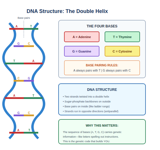
In 1953, James Watson and Francis Crick described the shape of DNA. They built on X-ray pictures of DNA made by Rosalind Franklin. DNA is shaped like a ladder that has been twisted along its length. This shape is called a double helix: “double” because there are two strands, and a helix is a spiral, like the thread of a screw.
The parts of DNA
The sugar-phosphate backbone. The two sides of the ladder are made of a sugar and a phosphate, taking turns: sugar, phosphate, sugar, phosphate, all the way along. The sugar is called deoxyribose. A phosphate is a small group of phosphorus and oxygen atoms. This backbone is strong, and it is the same all along the molecule.
The nitrogen bases. The rungs of the ladder are pairs of chemicals called bases. (They are called nitrogen bases, or nitrogenous bases, because they contain the element nitrogen.) DNA has four bases:
- Adenine (A) always pairs with Thymine (T)
- Guanine (G) always pairs with Cytosine (C)
This fixed pairing is called complementary base pairing. “Complementary” means the two fit together and complete each other. The two bases in a pair are held together by hydrogen bonds, which are weak pulls between the atoms of the two bases. An A-T pair has 2 hydrogen bonds, and a G-C pair has 3, so G-C pairs hold together slightly more strongly.
One strand of DNA reads A T G C. What does the other strand read, across from it?
Go one base at a time and use the pairing rule. Across from A is T. Across from T is A. Across from G is C. Across from C is G.
So the other strand reads T A C G. If you know one strand, you always know the other. That is the fact that lets DNA be copied, as Part 2 shows.
The genetic code
The order of the bases along the DNA is what carries the information. The 26 letters of the alphabet can spell a huge number of words, depending on their order. In the same way, the four bases of DNA can carry a huge amount of information, depending on their order.
A gene is a section of DNA that holds the instructions for making one particular protein. (Part 4 explains what proteins are and how they are built.) Humans have about 20,000 genes, but genes make up only about 2 percent of our DNA. Some of the rest controls when genes are turned on and off, and scientists are still working out what much of the rest does.
DNA is a double helix: two strands joined by pairs of bases. A always pairs with T, and G always pairs with C. The order of the bases is the code, and a gene is a section of that code.
Think it through. What are the four bases in DNA, and how do they pair?
Show a model answer
The four bases are adenine (A), thymine (T), guanine (G), and cytosine (C). A always pairs with T, and G always pairs with C. So if one strand has an A at some point, the other strand has a T across from it.
Think it through. Describe the basic structure of the DNA double helix.
Show a model answer
DNA is shaped like a twisted ladder. The two sides of the ladder are the sugar-phosphate backbones: sugar and phosphate, taking turns. The rungs are pairs of bases, A with T and G with C, held together by hydrogen bonds. The whole ladder is twisted into a spiral, which is the double helix.
How DNA is copied and packed
The GED test asks only the basic idea, which is this: before a cell divides, it makes a complete copy of its DNA, so that each new cell gets a full set of instructions. DNA is packed very tightly, and it is packed most tightly into chromosomes when a cell divides.
The details after this box go past the GED test. Read them if you want the whole story, or skip them on a first reading.
DNA replication
Before a cell divides, it must copy its DNA so that each new cell receives a complete set of genetic instructions. This copying is called DNA replication. The work is done by enzymes, which are proteins that make a chemical job happen quickly.
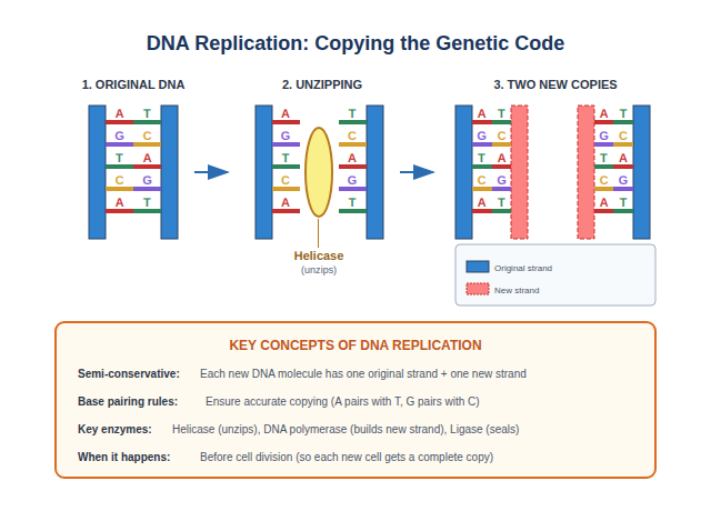
Step 1: Unzipping. The enzyme helicase separates the two strands by breaking the hydrogen bonds between the base pairs. The place where the strands come apart is shaped like a Y and is called the replication fork.
Step 2: Building new strands. The enzyme DNA polymerase reads each original strand and adds the matching bases to build a new partner strand. Because A pairs with T and G pairs with C, the order of the bases is copied exactly.
Step 3: Two identical copies. The result is two identical DNA molecules. Each one has one original strand and one new strand. This is called semi-conservative replication: “semi” means half, and “conservative” means kept. Half of each new molecule is the old strand, kept.
Why accurate copying matters
DNA replication is very accurate. After the checking and repairs are done, there is only about 1 mistake for every billion bases copied. This matters because a mistake in the DNA, called a mutation, can change a protein and can sometimes cause disease. The cell has several ways of catching and fixing mistakes:
- DNA polymerase checks its own work as it builds, and removes a base that does not match. This is called proofreading.
- Other enzymes, called mismatch repair enzymes, look over the newly copied DNA and fix mistakes that were missed.
- Even so, some mutations get through. Most make no difference. Some cause harm, and a few turn out to be helpful. Helpful mutations are one of the raw materials of evolution.
How DNA is packed: from molecule to chromosome
A single human cell holds about 6 feet of DNA, if you stretched it all out in a line. Yet it fits inside a nucleus only about 6 micrometers across. (A micrometer is one millionth of a meter, so the nucleus is far too small to see without a microscope.) This is possible because the DNA is packed very carefully:
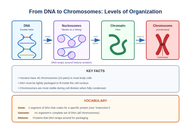
Nucleosomes. DNA wraps around small bundles of proteins called histones, the way thread wraps around a spool. One bundle of histones with DNA wrapped around it is called a nucleosome. Under a powerful microscope this looks like beads on a string: the nucleosomes are the beads, and the stretches of DNA between them are the string.
Chromatin. The nucleosomes coil into a thicker fiber called chromatin. For most of a cell’s life, its DNA is in this loosely packed form, so that the cell can get at the genes it needs.
Chromosomes. When the cell divides, the chromatin packs down even further into short, thick chromosomes. This tight packing protects the DNA while the cell divides, and it lets the DNA be sorted correctly into the new cells.
Think it through. What is the role of DNA polymerase in replication?
Show a model answer
DNA polymerase is the enzyme that builds the new strand. It reads an original strand one base at a time and adds the matching base: T across from A, A across from T, C across from G, and G across from C. It also checks its own work and removes a base that does not match.
Think it through. Why is DNA replication called “semi-conservative”?
Show a model answer
Because each new DNA molecule keeps (conserves) half of the old one. One of its two strands is an original strand, and the other is newly built. “Semi” means half.
Chromosomes, genes, and alleles
A chromosome is one long molecule of DNA, together with the proteins it is wrapped around. Each chromosome carries many genes.
Human chromosomes
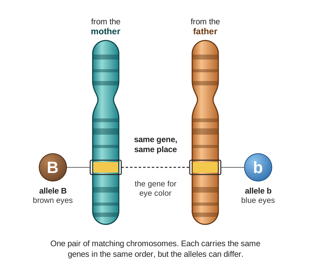
Most cells in the human body have 46 chromosomes, arranged in 23 pairs. One chromosome of each pair came from your mother, and one came from your father.
- Pairs 1 to 22 are called autosomes. They carry genes for traits of the body in general.
- Pair 23 is the sex chromosomes. Females usually have two X chromosomes (XX), and males usually have one X and one Y (XY).
Egg and sperm cells have only 23 chromosomes: one from each pair, which is one complete set. When an egg and a sperm join at fertilization, the new cell has 46 chromosomes: a complete set from each parent.
Because you have two copies of each chromosome, you have two copies of most genes, one from each parent. The two copies of a gene are not always the same. The different versions of a gene are called alleles, and Part 6 explains how they work.
Words from Parts 1 to 3
- DNA: deoxyribonucleic acid; the molecule that carries genetic information
- Gene: a section of DNA that holds the instructions for one protein
- Genome: all of the DNA of a living thing, the complete set
- Chromosome: one long, packed molecule of DNA with its proteins
- Histones: the proteins that DNA wraps around to be packed
- Replication: the copying of DNA before a cell divides
- Complementary base pairing: the rule that A pairs with T and G pairs with C
Think it through. How many chromosomes do humans have, and how are they arranged?
Show a model answer
Most human body cells have 46 chromosomes, in 23 pairs. In each pair, one chromosome came from the mother and one from the father. Pairs 1 to 22 are the autosomes. Pair 23 is the sex chromosomes: usually XX in females and XY in males. Egg and sperm cells have 23, one from each pair.
From gene to protein
The GED test asks only the basic idea, which is this: a gene holds the instructions for building a protein. The cell copies the gene’s message into a molecule called RNA, and structures called ribosomes read the RNA and build the protein. A mutation, a change in the DNA, can change the protein that gets built.
The details after this box go past the GED test. Read them if you want the whole story, or skip them on a first reading.
DNA holds instructions, but it does not build anything itself. The things that do most of the work in a cell are proteins. Proteins are large molecules made of a chain of smaller units called amino acids. Some proteins speed up chemical reactions, some form the structure of the body (such as the keratin in hair), and some carry messages or other molecules.
Using a gene’s instructions to build a protein is called gene expression. It takes two main steps, transcription and translation. Both steps use RNA (ribonucleic acid), a molecule much like DNA but with a single strand.
The central dogma
Genetic information flows along a set path, usually called the central dogma of molecular biology. (“Dogma” here just means a basic rule.)
In almost all living things, information flows this one way: DNA → RNA → protein. DNA stores the information, RNA carries the message, and proteins do the actual work of the cell.
It can help to picture the cell as a factory. The DNA is the master set of plans, which stays safe in the office (the nucleus). The mRNA is a working copy of one plan, sent out to the factory floor (the cytoplasm). The ribosome is the machine on the floor that reads the copy and builds the product. The protein is the finished product.
Step 1: Transcription (DNA → RNA)
Transcription is the copying of a gene’s information from DNA into messenger RNA, or mRNA. It happens in the nucleus. How it works:
- The enzyme RNA polymerase attaches to the start of the gene, at a stretch of DNA called the promoter, which acts as a start signal.
- RNA polymerase unwinds the DNA and reads one of its two strands, called the template strand.
- RNA polymerase builds the mRNA by adding the matching RNA building blocks, called nucleotides, one at a time.
- The finished mRNA comes off the DNA and leaves the nucleus through small openings called nuclear pores.
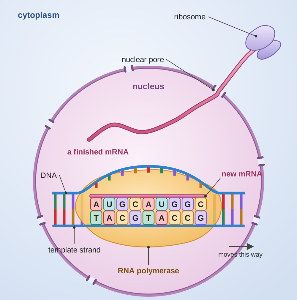
The key difference from DNA: RNA uses a base called uracil (U) in place of thymine (T). So where DNA would pair A with T, RNA pairs A with U.
The template strand of a short piece of DNA reads T A C G T A C C G. What mRNA is made from it?
Go one base at a time. Across from T, RNA puts A. Across from A, RNA puts U (not T). Across from C it puts G, and across from G it puts C.
| Strand | Bases |
|---|---|
| DNA template | T A C G T A C C G |
| mRNA made | A U G C A U G G C |
So the mRNA reads A U G C A U G G C.
Step 2: Translation (RNA → protein)
Translation is the reading of the mRNA to build a protein. It happens in the cytoplasm, the jelly-like inside of the cell, at small structures called ribosomes.
The genetic code: codons
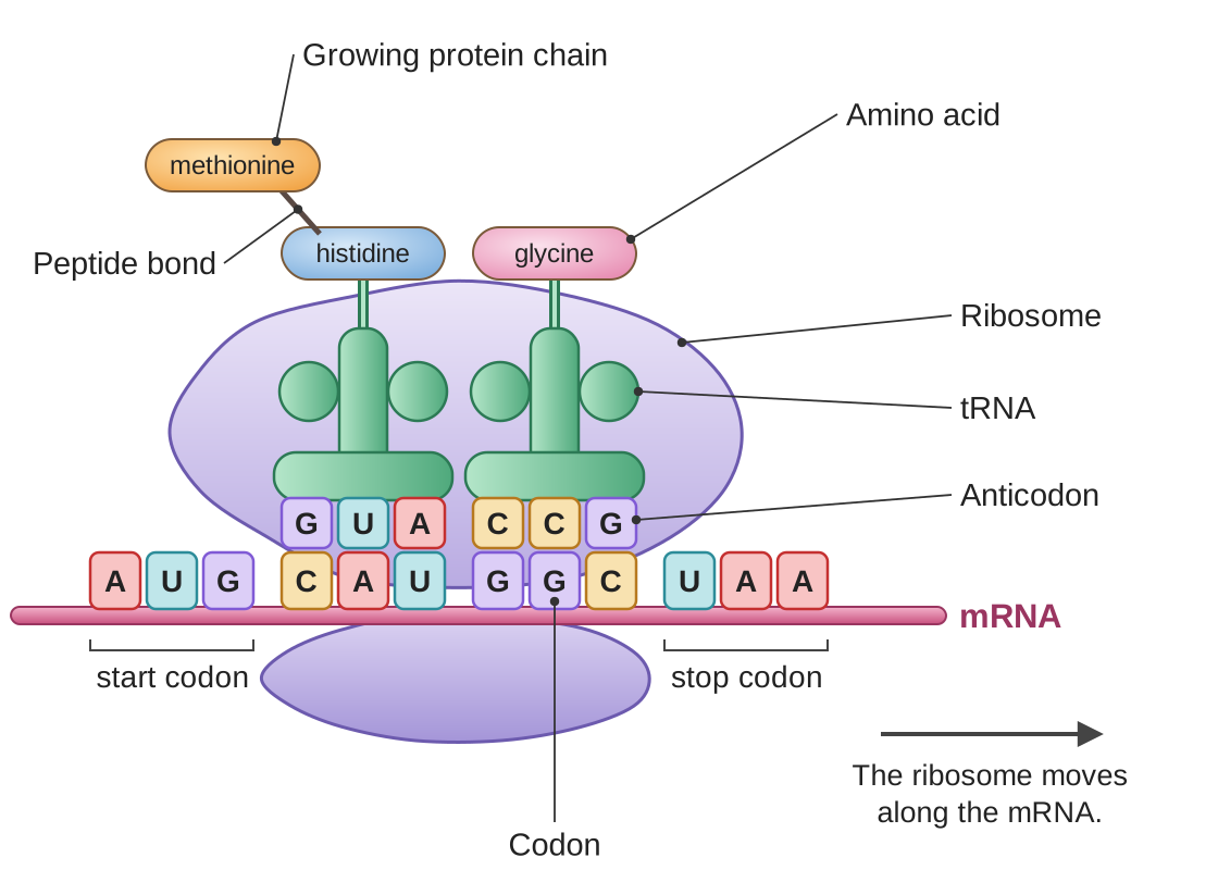
The mRNA is read three bases at a time. Each group of three bases is called a codon. Each codon stands for one amino acid, or for a stop signal. With 4 bases taken 3 at a time, there are 4 × 4 × 4 = 64 possible codons. That is more than enough for the 20 amino acids that proteins are made from.
- AUG is the start codon. It also stands for methionine, the first amino acid of the protein.
- UAA, UAG, and UGA are stop codons. They mark the end of the protein.
The genetic code is redundant: more than one codon can stand for the same amino acid. But it is not ambiguous: each codon stands for only one amino acid, never two.
An mRNA reads AUG CAU GGC UAA. What does the ribosome build?
| Codon | Means |
|---|---|
| AUG | start, and the amino acid methionine |
| CAU | the amino acid histidine |
| GGC | the amino acid glycine |
| UAA | stop |
The ribosome builds a short chain of three amino acids: methionine, then histidine, then glycine. At UAA it stops, and the chain is released.
Transfer RNA (tRNA)
Transfer RNA, or tRNA, is the molecule that matches each codon on the mRNA to the right amino acid:
- Each tRNA has an anticodon, a set of 3 bases that pairs with one particular codon on the mRNA.
- Each tRNA carries the one amino acid that goes with its anticodon.
- When the anticodon pairs with the codon, the tRNA hands its amino acid over to the growing protein chain.
Putting it together: how translation works
- The ribosome attaches to the mRNA at the start codon, AUG.
- A tRNA with the matching anticodon, UAC, brings the amino acid methionine.
- The ribosome moves to the next codon, and another tRNA brings the next amino acid.
- The amino acids are joined together by chemical links called peptide bonds.
- This goes on until the ribosome reaches a stop codon.
- The finished protein is released and folds into the shape it needs to do its job.
Why the shape of a protein matters
A protein is not just a chain. It folds into a particular three-dimensional shape, and the order of its amino acids decides that shape. The shape decides what the protein can do:
- An enzyme must fit the molecule it works on the way a key fits a lock.
- An antibody, a protein of the immune system, must fit a particular shape on a germ in order to recognize it.
- A receptor, a protein that receives signals, must fit a particular hormone or other signaling molecule.
If a mutation changes an amino acid, it may change the protein’s shape, and so change what the protein can do. Many genetic diseases happen this way.
Mutations and proteins
A mutation is any change in the order of the bases in DNA. A mutation can affect a protein in different ways:
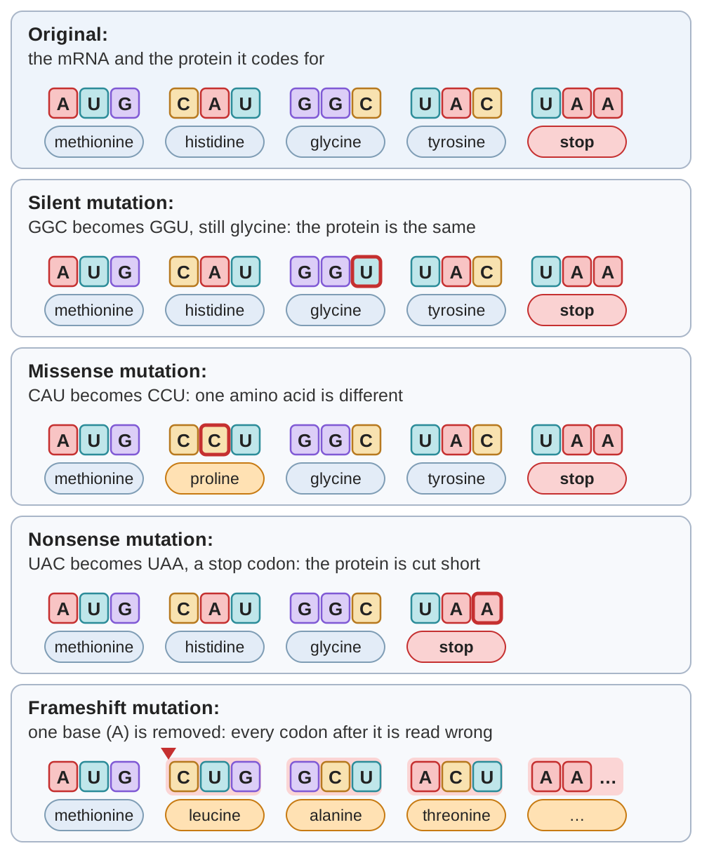
- Silent mutation: a codon changes, but it still stands for the same amino acid (because the code is redundant). The protein is not changed.
- Missense mutation: a codon changes to one that stands for a different amino acid. This may or may not change how the protein works, depending on where the change is and how different the new amino acid is.
- Nonsense mutation: a codon changes to a stop codon, so the protein is cut off too early. The protein usually does not work.
- Frameshift mutation: one or more bases are added or removed (not in a multiple of 3). Because the mRNA is read in threes, every codon after that point is read wrong. The protein is usually badly damaged.
Think it through. What are the two main steps of gene expression, and where does each happen?
Show a model answer
The first step is transcription: a gene’s DNA is copied into mRNA. It happens in the nucleus. The second step is translation: the mRNA is read and a protein is built. It happens in the cytoplasm, at the ribosomes.
Think it through. What is a codon? What is an anticodon?
Show a model answer
A codon is a group of three bases on the mRNA that stands for one amino acid, or for stop. An anticodon is the set of three bases on a tRNA that pairs with a codon. For example, the codon AUG pairs with the anticodon UAC.
Think it through. What job does tRNA do in translation?
Show a model answer
tRNA brings the right amino acid to the ribosome. Its anticodon pairs with a codon on the mRNA, and it carries the amino acid that goes with that codon, so the amino acids are added to the chain in the order the mRNA calls for.
Think it through. Why does the shape of a protein matter for what it does?
Show a model answer
A protein works by fitting other molecules, the way a key fits a lock: an enzyme fits the molecule it acts on, an antibody fits a germ, a receptor fits a hormone. If the shape changes, the protein may no longer fit, and then it cannot do its job.
Think it through. What is a frameshift mutation, and why is it usually harmful?
Show a model answer
A frameshift mutation adds or removes bases in a number that is not a multiple of 3. Because the mRNA is read three bases at a time, every codon after the change is read wrong, so every amino acid after that point can be wrong, and a stop codon may appear too early. The protein that results usually cannot work.
Cell division: mitosis and meiosis
Cells make more cells by dividing. But not all cell division is the same. Your body uses two different kinds: mitosis, for growth and repair, and meiosis, for making sex cells. The difference between them is the key to understanding inheritance.
The study guide Cell Division covers this subject at length, stage by stage. This part keeps what genetics needs.
Why cells divide
- Growth: a baby grows into an adult by making more cells.
- Repair: cuts heal, and damaged tissue is replaced.
- Reproduction: living things make offspring.
Before any cell divides, it must first copy its DNA, so that each new cell gets a complete set of instructions.
Counting chromosomes: diploid and haploid
Before looking at mitosis and meiosis, you need two words for how many chromosomes a cell has:
- Diploid (2n): having two complete sets of chromosomes, one set from each parent. Most human cells are diploid, with 46 chromosomes (23 pairs).
- Haploid (n): having one complete set of chromosomes. Human eggs and sperm, called gametes, are haploid, with 23 chromosomes.
When an egg and a sperm join at fertilization: n + n = 2n, or 23 + 23 = 46.
Mitosis: division for growth and repair
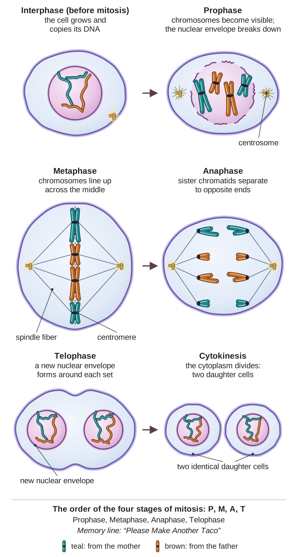
Mitosis makes two genetically identical cells, called daughter cells, from one parent cell. It is used for growth, for repair, and by some living things to reproduce without a partner (asexual reproduction).
The stages of mitosis (PMAT)
Interphase (before mitosis): The cell grows, does its normal work, and copies its DNA. Each chromosome is now two identical copies, called sister chromatids, joined at a point called the centromere.
Prophase: The chromosomes pack down and become visible. The envelope around the nucleus starts to break down. Thin fibers, called spindle fibers, form.
Metaphase: The chromosomes line up across the middle of the cell. Spindle fibers attach at the centromeres. This is the best stage for photographing chromosomes.
Anaphase: The sister chromatids separate and move to opposite ends of the cell. The cell gets longer.
Telophase: A new nuclear envelope forms around each set of chromosomes. The chromosomes loosen again.
Cytokinesis: The cytoplasm divides, making two separate daughter cells. In animal cells, the cell pinches in two along a groove called the cleavage furrow.
Meiosis: division for sexual reproduction
Meiosis makes four haploid cells, the gametes, from one diploid parent cell, and each of the four is genetically different. It takes two divisions.
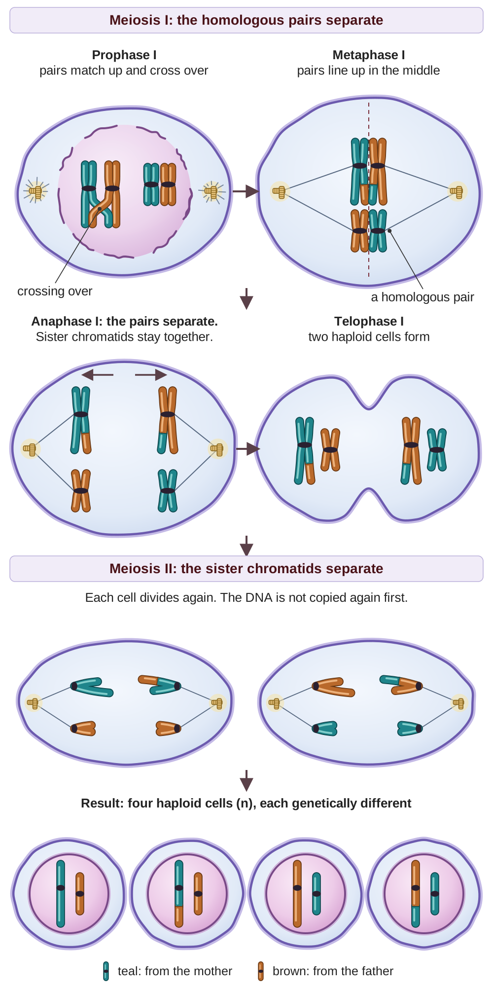
Meiosis I: separating the matching pairs
In meiosis I, the homologous chromosomes are separated. Homologous chromosomes are a matching pair: one from the mother, one from the father, carrying the same genes.
- Prophase I: The homologous chromosomes pair up and trade sections with each other. This is called crossing over. It shuffles genes between the chromosome from the mother and the one from the father, making new combinations.
- Metaphase I: The homologous pairs line up in the middle. Which chromosome of a pair ends up on which side is a matter of chance. This is called independent assortment.
- Anaphase I: The homologous chromosomes separate. The sister chromatids stay together.
- Telophase I and cytokinesis: Two haploid cells form, each with one chromosome from each pair.
Meiosis II: separating the sister chromatids
Meiosis II works much like mitosis: the sister chromatids are separated.
- The DNA is not copied again between meiosis I and meiosis II.
- Each of the two cells divides again.
- The sister chromatids separate and move to opposite ends.
- The result is four haploid cells, each genetically different.
Where the variety comes from
Meiosis makes each sex cell genetically different in two ways:
- Crossing over: In prophase I, homologous chromosomes trade sections. This makes chromosomes with new combinations of alleles that did not exist in either parent.
- Independent assortment: In metaphase I, each homologous pair lines up by chance. With 23 pairs, this alone gives 223 possible combinations, which is more than 8 million.
Add the variety from the other parent, and the chance of which sperm meets which egg, and the number of possible combinations is enormous. That is why brothers and sisters are genetically different from each other, except for identical twins.
Comparing mitosis and meiosis
| Mitosis | Meiosis | |
|---|---|---|
| Purpose | growth, repair, replacing cells | making sex cells (eggs and sperm) |
| Number of divisions | one | two |
| Result | 2 identical cells, each with two sets (2n) | 4 different cells, each with one set (n) |
| Genetic variation | none: the new cells are copies of the parent cell | high: each sex cell is different |
| Where it happens | body cells, all through the body | only in the reproductive organs (ovaries and testes) |
| Crossing over | no | yes: it shuffles alleles |
Going further · When chromosomes fail to separate: nondisjunction
The GED test asks only the basic idea, which is this: sometimes chromosomes do not separate correctly during meiosis, so an egg or sperm has one chromosome too many or too few. Down syndrome is caused by an extra copy of chromosome 21.
The details after this box go past the GED test. Read them if you want the whole story, or skip them on a first reading.
Nondisjunction happens when chromosomes fail to separate correctly during meiosis. The result is a gamete with too many or too few chromosomes.
If a gamete with an extra chromosome is fertilized, the child has trisomy: three copies of that chromosome instead of two. If a gamete that is missing a chromosome is fertilized, the result is monosomy: only one copy.
Down syndrome (trisomy 21), three copies of chromosome 21, is the most common example among babies who are born. Most other trisomies and monosomies of the autosomes end in miscarriage. An extra or missing sex chromosome is usually survivable: for example, Turner syndrome (only one X) and Klinefelter syndrome (XXY).
Words from Part 5
- Diploid (2n): two complete sets of chromosomes (46 in humans)
- Haploid (n): one complete set of chromosomes (23 in humans)
- Sister chromatids: the two identical copies of a chromosome, joined at the centromere
- Homologous chromosomes: a matching pair, one from each parent
- Crossing over: the trading of sections between homologous chromosomes
- Independent assortment: the chance sorting of chromosomes into sex cells during meiosis
- Nondisjunction: the failure of chromosomes to separate correctly
Think it through. What is the difference between diploid and haploid?
Show a model answer
A diploid cell has two complete sets of chromosomes, one from each parent; in humans that is 46. A haploid cell has one complete set; in humans that is 23. Body cells are diploid, and eggs and sperm are haploid.
Think it through. What are the four stages of mitosis?
Show a model answer
Prophase, metaphase, anaphase, and telophase: PMAT. (Interphase comes before, when the DNA is copied, and cytokinesis comes at the end, when the cell splits.)
Think it through. How many cells come from mitosis? From meiosis? Are they identical or different?
Show a model answer
Mitosis makes 2 cells, identical to each other and to the parent cell. Meiosis makes 4 cells, each with one set of chromosomes, and each genetically different.
Think it through. What are two sources of genetic variety in meiosis?
Show a model answer
Crossing over, when homologous chromosomes trade sections in prophase I; and independent assortment, when each pair lines up by chance in metaphase I, so each sex cell gets a different mix of chromosomes from the mother and the father.
Think it through. What is nondisjunction, and what can it cause?
Show a model answer
Nondisjunction is when chromosomes fail to separate correctly during meiosis, so a sex cell ends up with an extra chromosome or a missing one. It can cause a trisomy, such as Down syndrome (three copies of chromosome 21), or a monosomy, such as Turner syndrome (one X).
Inheritance: dominant and recessive
How are traits passed from parents to their children? Gregor Mendel, a monk who lived in the 1800s, worked out the basic rules by breeding pea plants and counting the traits of their offspring. His rules are still the foundation of genetics today.
Mendel’s main discoveries
- Traits are decided by separate units. Today we call these units genes. Each parent passes one copy of each gene, called an allele, to each child.
- Some alleles are dominant, and others are recessive. A dominant allele hides a recessive allele when both are present.
- The two alleles separate when sex cells form. This is the Law of Segregation. Each egg or sperm gets only one allele of each gene.
- Different genes are sorted separately. This is the Law of Independent Assortment. Which allele you get for one gene does not affect which you get for another. (There are exceptions, for genes that sit close together on the same chromosome.)
Words for inheritance
- Gene: a section of DNA that holds the instructions for a protein, and so affects a trait
- Allele: one version of a gene, such as the allele for purple flowers or the allele for white flowers
- Genotype: the alleles a living thing has, written as letters, such as Bb
- Phenotype: the trait that actually shows, what you can see or measure, such as brown eyes
- Homozygous: having two identical alleles, such as BB or bb
- Heterozygous: having two different alleles, such as Bb
Dominant and recessive alleles
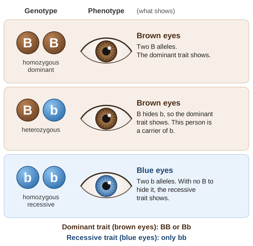
When genotypes are written, a dominant allele is written with a capital letter, and a recessive allele with the same letter in lowercase. Take B for brown eyes and b for blue eyes:
- BB is homozygous dominant. The dominant trait shows: brown eyes.
- Bb is heterozygous. The dominant trait shows (brown eyes), but the person carries the recessive allele.
- bb is homozygous recessive. The recessive trait shows: blue eyes.
(Real eye color is shaped by several genes. Textbooks often treat it as one gene with two alleles to keep the example simple.)
A carrier is a person who is heterozygous for a recessive trait. The carrier does not show the trait but can pass the recessive allele on to children. For example, cystic fibrosis, a disease of the lungs and digestion, is caused by a recessive allele. A person with one normal allele and one cystic fibrosis allele is healthy but is a carrier.
Think it through. What is the difference between genotype and phenotype?
Show a model answer
The genotype is the pair of alleles a living thing has, written in letters, such as Bb. The phenotype is the trait that actually shows, such as brown eyes. Two different genotypes can give the same phenotype: BB and Bb both show brown eyes.
Think it through. What is a carrier?
Show a model answer
A carrier is a person who has one dominant allele and one recessive allele for a trait (heterozygous). The dominant allele hides the recessive one, so the carrier does not show the trait, but the carrier can pass the recessive allele to a child.
Punnett squares and probability
A Punnett square is a small grid for predicting what the offspring of two parents might inherit. It shows every possible combination of the parents’ alleles, and each box is equally likely. The GED test gives Punnett squares often and asks you to read them.
How to use a Punnett square
- Write one parent’s two alleles across the top, and the other parent’s two alleles down the side.
- Remember that each parent gives only ONE allele to each child.
- Fill in each box with the letter from the top of its column and the letter from the side of its row.
- Count the boxes to find the ratios. Each box is 1 out of 4, or 25 percent.
For a cross between two heterozygous parents (Aa × Aa):
- Genotype ratio: 1 AA : 2 Aa : 1 aa
- Phenotype ratio: 3 dominant : 1 recessive (3:1)
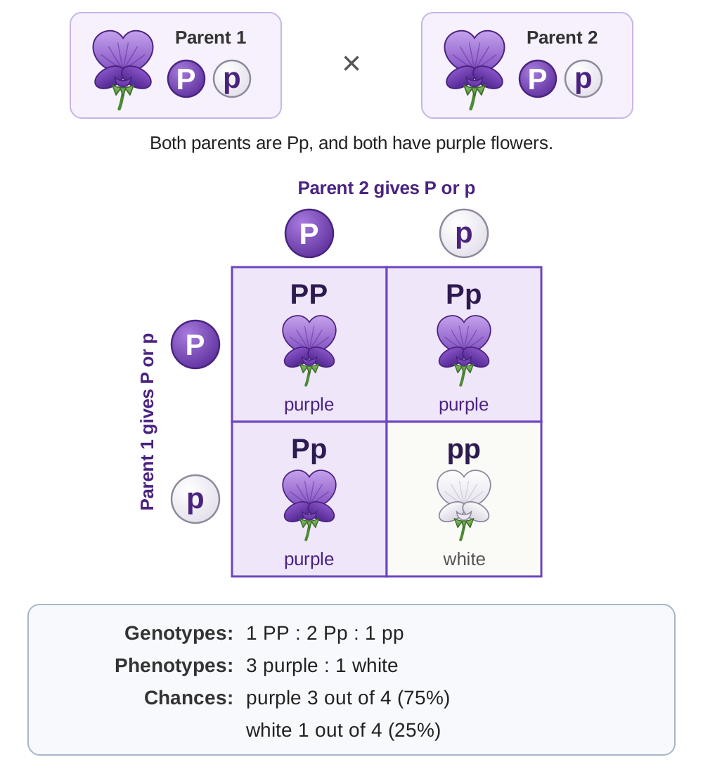
In pea plants, purple flowers (P) are dominant over white flowers (p). Two Pp plants are crossed. What fraction of the offspring are likely to have white flowers?
Step 1. Put one parent’s alleles, P and p, across the top. Put the other parent’s, P and p, down the side.
Step 2. Fill each box with one letter from its column and one from its row:
| P | p | |
|---|---|---|
| P | PP | Pp |
| p | Pp | pp |
Step 3. Count. Only pp shows the recessive trait, white. That is 1 box out of 4.
Answer: 1 out of 4, or 25 percent, of the offspring are likely to have white flowers. The other 3 out of 4 (75 percent) have purple flowers: the PP box and the two Pp boxes.
Notice that two purple parents can have a white-flowered offspring, because each parent carries a hidden p.
Two parents are both carriers of cystic fibrosis. Call the normal allele F and the cystic fibrosis allele f, so each parent is Ff. What is the chance that their child will have cystic fibrosis? What is the chance the child will be a carrier?
| F | f | |
|---|---|---|
| F | FF | Ff |
| f | Ff | ff |
Only ff has the disease. That is 1 box out of 4: a 25 percent chance, or 1 in 4.
The two Ff boxes are carriers: 2 out of 4, a 50 percent chance.
The FF box is neither sick nor a carrier: 1 out of 4, 25 percent.
One more point about chance. The 1-in-4 chance holds for each child, separately. If this couple already has three healthy children, the chance that the fourth child has cystic fibrosis is still 1 in 4. The square gives the chances for each birth; it does not promise that 1 child in every 4 will have the disease.
Practice: Punnett squares
Choose an answer, then press Check. The explanation opens either way.
Brown (B) is dominant over blue (b). A parent who is Bb has a child with a parent who is bb. What is the chance the child is bb?
The square has Bb, Bb, bb, bb: two boxes out of four are bb, so the chance is 2 out of 4, or 50 percent. If you chose 1 out of 4, you may have used the answer for two Bb parents; here one parent is bb, so half the boxes get a b from both.
A PP plant (purple) is crossed with a pp plant (white). What will the offspring look like?
Every box gets a P from the first parent and a p from the second, so every offspring is Pp, and all of them show the dominant trait, purple. The 3-to-1 ratio belongs to a cross of two Pp parents, not this one.
Two carriers (Ff) already have three children, and none of them has cystic fibrosis. What is the chance that their fourth child will have it?
Each child is a new, separate chance, and for each one the chance of ff is 1 out of 4. The earlier children do not change it. The tempting mistake is to think the square promises one sick child in every four.
Think it through. If both parents are heterozygous (Aa), what is the chance their child will show the recessive trait?
Show a model answer
1 out of 4, or 25 percent. The Punnett square gives AA, Aa, Aa, and aa, and only aa shows the recessive trait.
Blood type, sex-linked traits, and other patterns
Not every trait follows the simple dominant-and-recessive pattern. This part covers four other patterns, at the level the GED test asks about.
Beyond simple dominance
Incomplete dominance: neither allele fully hides the other, so a heterozygous offspring shows a blend of the two traits. Example: in snapdragon flowers, a red-flowered plant crossed with a white-flowered plant gives pink flowers.
Codominance: both alleles show fully, side by side, in a heterozygous person. Example: in blood type AB, the red blood cells carry both the A marker and the B marker. (These markers are molecules on the surface of the red blood cell, called antigens.)
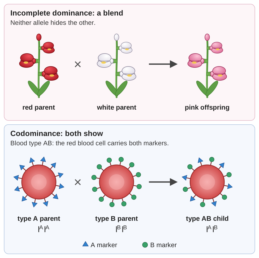
Multiple alleles: a gene has more than two alleles in the population, though each person still has only two. Example: the blood type gene has three alleles, written IA, IB, and i.
Blood type: a classic example
Blood type shows both codominance and multiple alleles. IA and IB are codominant: when both are present, both show, and the blood type is AB. The allele i is recessive to both. Here is every genotype and the blood type it gives:
| Blood type | Genotypes that give it | Notes |
|---|---|---|
| Type A | IAIA or IAi | homozygous or heterozygous |
| Type B | IBIB or IBi | homozygous or heterozygous |
| Type AB | IAIB | both A and B show: codominance |
| Type O | ii | only when both alleles are the recessive i |
A parent with type A blood (IAi) and a parent with type B blood (IBi) have a child. What blood types are possible?
| IB | i | |
|---|---|---|
| IA | IAIB | IAi |
| i | IBi | ii |
The four boxes give type AB (IAIB), type A (IAi), type B (IBi), and type O (ii). Each has a 25 percent chance. So these two parents could have a child of any of the four blood types. A type O child is possible because each parent carries a hidden i.
Sex-linked traits
Some genes sit on the sex chromosomes, X and Y. A trait controlled by a gene on the X chromosome is called X-linked, or sex-linked. Such traits are passed on differently in males and females:
- Males have only one X chromosome (XY). So a male shows whatever allele is on his one X.
- Females have two X chromosomes (XX). So a recessive allele on one X can be hidden by a dominant allele on the other.
That is why conditions such as red-green color blindness and hemophilia (a disorder in which the blood does not clot properly) are more common in males. A male needs only one copy of the recessive allele to have the condition, while a female needs two.
A woman is a carrier for color blindness (XBXb, where B is normal color vision and b is color blindness). Her partner has normal color vision (XBY). What are the chances for their children?
| XB | Y | |
|---|---|---|
| XB | XBXB | XBY |
| Xb | XBXb | XbY |
The mother passes her color-blindness allele to each child half the time (a 50 percent chance). What that means depends on whether the child is a son or a daughter:
- A son gets his X from his mother and his Y from his father. Half of the sons are expected to get Xb, and a son with XbY is color blind.
- A daughter gets an X from each parent. Her father’s XB hides the b, so a daughter who gets Xb from her mother is a carrier (XBXb), not color blind.
So each son has a 50 percent chance of being color blind, and each daughter has a 50 percent chance of being a carrier. None of the daughters is expected to be color blind.
Traits controlled by many genes
Many traits are controlled by several genes working together. This is called polygenic inheritance (“poly” means many). Such traits do not fall into a few separate types. Instead, they vary smoothly across a whole range. Examples:
- Height
- Skin color
- Intelligence
- The risk of many diseases
These traits are also shaped by the environment, such as food, health care, and living conditions. So their inheritance is complicated, and a simple Punnett square cannot predict them.
Think it through. How is codominance different from simple dominance?
Show a model answer
In simple dominance, the dominant allele hides the recessive one, so a heterozygous person shows only the dominant trait. In codominance, neither allele hides the other: both show fully. A person with type AB blood shows both the A marker and the B marker.
Think it through. Why are X-linked recessive conditions more common in males?
Show a model answer
Males have only one X chromosome, so a single recessive allele on it is enough to cause the condition; there is no second X to hide it. Females have two X chromosomes, so they need the recessive allele on both to have the condition. With one copy, a female is a carrier.
Genetic technology: DNA fingerprinting, genetic engineering, and CRISPR
The GED test asks only the basic idea, which is this: scientists can compare people’s DNA to identify them (DNA fingerprinting); they can move a gene from one living thing into another, so that, for example, bacteria make human insulin (genetic engineering); and a newer tool called CRISPR lets them edit a gene directly.
The details after this box go past the GED test. Read them if you want the whole story, or skip them on a first reading.
New genetic tools have changed medicine, farming, and the criminal courts. Knowing how they work helps you make sense of news about genetic testing, genetically modified foods, and gene therapy (treating a disease by changing a person’s genes).
DNA fingerprinting
DNA fingerprinting, also called DNA profiling, identifies a person by the pattern in his or her DNA. More than 99 percent of human DNA is the same from one person to the next, but certain regions differ enough to tell people apart.
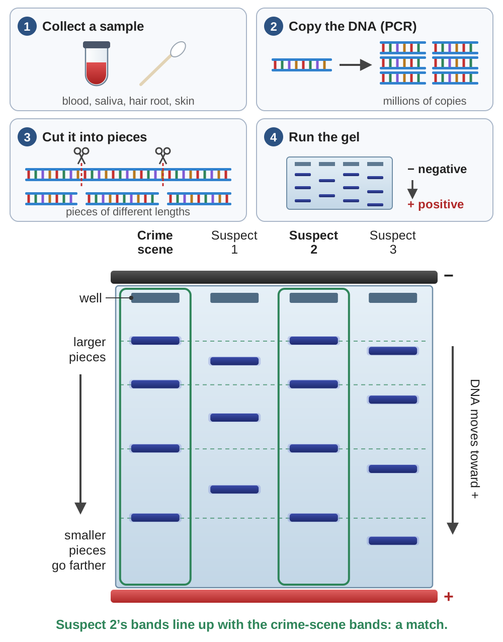
How DNA fingerprinting works
- Collect a DNA sample: blood, saliva, the root of a hair, skin cells, or other material from the body.
- Take out the DNA and make copies: a method called PCR (polymerase chain reaction) makes millions of copies of chosen regions of the DNA, so that even a tiny sample can be studied.
- Cut the DNA with restriction enzymes: these enzymes cut DNA wherever a particular short sequence of bases appears, leaving pieces of different lengths. (This was the step in the original method. Most labs today skip it: they use PCR to copy short, repeating regions whose length differs from person to person, and then measure those lengths.)
- Separate the pieces by gel electrophoresis: the pieces are placed in a gel, and an electric current is run through it. DNA carries a negative charge, so it moves toward the positive end. Smaller pieces move faster and travel farther.
- Look at the pattern and compare: the pattern of bands is different for every person, except identical twins.
Uses of DNA fingerprinting
- Criminal investigations: matching DNA from a crime scene to a suspect.
- Paternity testing: finding a child’s father. A child inherits half of its DNA from each parent, so half of the child’s bands must match the father’s.
- Identifying remains: naming the victims of disasters, and in historical investigations.
- Freeing the innocent: DNA evidence has freed many people who were convicted of crimes they did not commit.
Genetic engineering and recombinant DNA
Genetic engineering means changing a living thing’s DNA directly, to add, remove, or change genes. Recombinant DNA is DNA that combines genetic material from different sources, such as a human gene placed inside bacterial DNA.
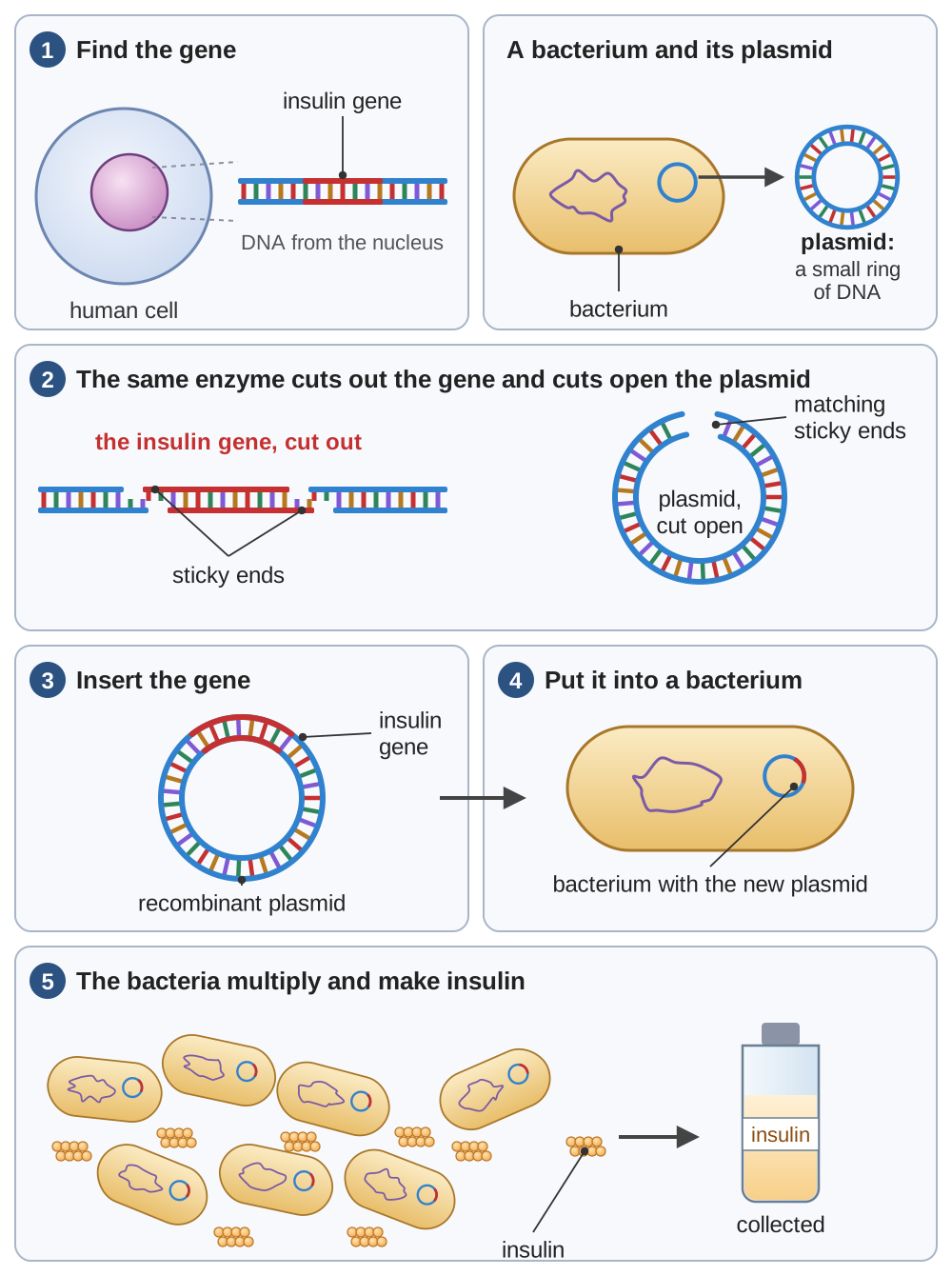
The basic process
- Identify the gene you want: for example, the human gene for insulin.
- Cut the gene out: restriction enzymes cut the DNA at particular sequences. The cuts leave short single-stranded tails, called sticky ends, that can pair with matching tails.
- Put the gene into a carrier: the gene is inserted into a plasmid (a small ring of DNA found in bacteria) or into a virus that can carry it into cells. The carrier is called a vector. The same restriction enzyme is used to cut the plasmid open, so its sticky ends match the gene’s.
- Put the plasmid into host cells: the recombinant plasmid is taken into bacteria or other cells. This step is called transformation.
- Select and grow: the cells that took in the plasmid are picked out and grown in large numbers.
- Collect the product: the protein made from the inserted gene is collected.
Products of genetic engineering
Medical products:
- Insulin: the first medicine made with recombinant DNA (approved in 1982). Over time it replaced insulin taken from pigs and cows.
- Human growth hormone: for treating some growth disorders.
- Clotting factors: the proteins that help blood clot, for people with hemophilia.
- Vaccines: the hepatitis B vaccine is made with recombinant DNA.
Farm products, often called GMOs (genetically modified organisms):
- Pest-resistant crops: Bt corn carries a gene from a bacterium (Bacillus thuringiensis), so the corn makes a protein that kills certain insects that eat it.
- Herbicide-tolerant crops: these survive weed-killing sprays, so farmers can kill the weeds without harming the crop.
- Golden rice: engineered to make beta-carotene, which the body turns into vitamin A.
- Drought-resistant crops: able to grow with less water, which matters more as the climate changes.
CRISPR: a tool for editing genes
CRISPR-Cas9 (say CRISP-er cass-NINE) is a newer tool that lets scientists change DNA at an exact spot. It is faster, cheaper, and more precise than earlier methods.
How CRISPR works
- Design a guide RNA: a short piece of RNA is made to match the stretch of DNA the scientists want to change.
- The guide RNA finds the target: it leads a protein called Cas9 to that exact place in the DNA.
- Cas9 cuts the DNA: Cas9 cuts through both strands, like a pair of scissors made of a molecule.
- The cell repairs the cut: the cell’s own repair system fixes the break. If the repair is sloppy, the gene stops working, which is one way to switch a gene off. If the scientists supply a piece of DNA to copy from, the cell can use it to write in a new sequence.
What CRISPR is used for, and may be used for
- Treating genetic diseases: such as sickle cell disease, muscular dystrophy, and some kinds of blindness. In 2023, a CRISPR treatment for sickle cell disease became the first to be approved in the United States.
- Cancer research and treatment: changing a patient’s immune cells so that they attack cancer.
- Farming: changing crops more precisely than older genetic engineering methods.
- Basic research: learning what genes do, by switching them off one at a time.
Think it through. How does gel electrophoresis separate pieces of DNA?
Show a model answer
The pieces of DNA are placed in a gel, and an electric current is run through it. DNA has a negative charge, so it moves toward the positive end. Small pieces slip through the gel faster and travel farther; large pieces move slowly and stay nearer the start. So the pieces end up sorted by size, as bands.
Think it through. What is a plasmid, and why is it useful in genetic engineering?
Show a model answer
A plasmid is a small ring of DNA found in bacteria, separate from the bacterium’s main DNA. It is useful because scientists can cut it open, put a gene into it, and get bacteria to take it in. The bacteria then copy the plasmid as they multiply and make the protein the gene codes for, such as human insulin.
Think it through. Name two medical products made with recombinant DNA.
Show a model answer
Any two of these: insulin, human growth hormone, clotting factors for hemophilia, and the hepatitis B vaccine.
Think it through. What does CRISPR-Cas9 do, and why does it matter?
Show a model answer
CRISPR-Cas9 cuts DNA at an exact spot chosen by a guide RNA, so that a gene can be switched off or changed. It matters because it is faster, cheaper, and more precise than earlier methods, and it can be used to treat genetic diseases such as sickle cell disease, as well as in cancer treatment, farming, and research.
Genetics in society: ethics and privacy
Genetic technology raises questions that are not only scientific. They are questions about fairness, safety, and privacy, and a society has to decide them together. The GED test may ask you to weigh an argument about them, so it helps to know the main issues.
Questions of right and wrong
Editing the genes that are passed on: Should we edit human embryos? Changes made to an embryo, or to eggs or sperm, would be passed on to all of that person’s descendants. (This is called germline editing.) In 2018, a scientist in China announced that he had made the first gene-edited babies, and scientists around the world condemned it.
Access and fairness: Who will benefit from gene treatments? Will only wealthy people be able to pay for them?
Unexpected effects: What if an edit has effects nobody expected? Scientists do not yet fully understand how all genes work together.
Enhancement or treatment: Is it acceptable to edit genes not to treat a disease but to make someone “better”, for example taller or stronger?
Concerns about GMOs: effects on the environment, a few large companies controlling the food supply, and whether GMO foods should be labeled.
Genetic testing and privacy
Genetic testing can reveal a person’s risk of disease, his or her ancestry, and other personal information. That raises questions of privacy:
- Who should be allowed to see your genetic information?
- Can insurance companies or employers use your genetic information against you?
- What happens to the DNA data from home DNA tests you buy online?
A federal law, the Genetic Information Nondiscrimination Act (GINA), passed in 2008, gives some protection. It forbids health insurers from using your genetic information to deny you coverage or to set your price. It also forbids employers with 15 or more workers from using your genetic information to decide whether to hire, fire, pay, or promote you. But GINA does not cover life insurance, disability insurance, or long-term care insurance. Those companies may still ask about genetic test results.
Think it through. What is one ethical concern about gene-editing technology?
Show a model answer
Any one of these, explained: Germline editing, because changes to an embryo would be passed on to future generations who never agreed to them. Fairness, because the treatments may be so costly that only wealthy people can get them. Unexpected effects, because we do not fully understand how genes work together. Enhancement, because editing genes to make people taller or stronger, rather than to treat disease, raises the question of who decides what counts as “better.”
Think it through. A worker is offered a job and is thinking about taking a home DNA test. What does GINA protect, and what does it not protect?
Show a model answer
GINA says the employer (if it has 15 or more workers) may not use the worker’s genetic information in hiring, firing, pay, or promotion, and a health insurer may not use it to deny coverage or raise the price. GINA does not cover life insurance, disability insurance, or long-term care insurance, which may ask about genetic test results. GINA also does not control what the DNA testing company itself does with the data; that depends on the company’s own rules, so it is worth reading them first.
The terms in this guide
Allele one version of a gene, such as the allele for purple flowers or for white flowers.
Base one of the four chemicals (A, T, G, C) that make the rungs of DNA. RNA uses U in place of T.
Carrier a person with one dominant and one recessive allele who does not show the recessive trait but can pass it on.
Chromosome one long, packed molecule of DNA with its proteins. Humans have 46, in 23 pairs.
Codominance both alleles show fully in a heterozygous person, as in type AB blood.
Diploid having two complete sets of chromosomes (46 in humans).
DNA deoxyribonucleic acid, the molecule that carries genetic information.
Dominant an allele that shows even when only one copy is present; written with a capital letter.
Double helix the shape of DNA: two strands twisted like a spiral ladder.
Gene a section of DNA that holds the instructions for one protein.
Genotype the alleles a living thing has, written as letters, such as Bb.
GINA the Genetic Information Nondiscrimination Act (2008), which protects against the use of genetic information by health insurers and employers.
Haploid having one complete set of chromosomes (23 in humans), as eggs and sperm do.
Heterozygous having two different alleles, such as Bb.
Homozygous having two identical alleles, such as BB or bb.
Incomplete dominance neither allele fully hides the other, so the trait is a blend, as with pink snapdragons.
Meiosis cell division that makes four different sex cells, each with one set of chromosomes.
Mitosis cell division that makes two identical cells, each with two sets of chromosomes.
Mutation any change in the order of the bases in DNA.
Phenotype the trait that actually shows, such as brown eyes or white flowers.
Protein a large molecule, built from a chain of amino acids, that does much of the work in a cell.
Punnett square a grid that shows every possible combination of two parents’ alleles, each equally likely.
Recessive an allele that shows only when two copies are present; written with a lowercase letter.
RNA ribonucleic acid, a one-stranded molecule like DNA that carries a gene’s message to the ribosomes.
Sex-linked trait a trait controlled by a gene on the X chromosome, such as red-green color blindness.
12 questions on this guide
Check yourself
Choose an answer, then press Check. The explanation opens either way.
One strand of DNA reads A T T G C. What does the other strand read, across from it?
A pairs with T, and G pairs with C, so A T T G C is matched by T A A C G. The choice with U is a trap: uracil (U) is found in RNA, not DNA.
Most human body cells have 46 chromosomes. How many chromosomes does a human sperm cell have?
A sperm cell is haploid: it has one chromosome from each of the 23 pairs, which is one complete set of 23. The common mistake is 46, which is the number in body cells, not in sex cells.
For eye color in this example, B (brown) is dominant and b (blue) is recessive. A person’s genotype is Bb. What is this genotype called?
Bb has two different alleles, so it is heterozygous. It is tempting to call it homozygous dominant because it has a capital B, but homozygous dominant means two capitals, BB.
Two parents are both Aa. What is the chance that their child shows the recessive trait?
The Punnett square gives AA, Aa, Aa, and aa. Only aa shows the recessive trait: 1 box out of 4, or 25 percent. 75 percent is the chance of showing the dominant trait, which is easy to confuse with the question.
Cystic fibrosis is caused by a recessive allele, f. Which person is a carrier?
A carrier has one dominant and one recessive allele, Ff: the disease is hidden, but the f can be passed on. The person who is ff is not a carrier; that person has the disease.
A parent with blood type AB (IAIB) has a child with a parent with blood type O (ii). Which blood types can the child have?
The AB parent passes either IA or IB, and the O parent always passes i. So the child is IAi (type A) or IBi (type B). It is tempting to pick the parents’ own types, AB or O, but no child can get two i’s or both IA and IB.
A red snapdragon is crossed with a white snapdragon, and all the offspring have pink flowers. What pattern of inheritance is this?
Pink is a blend of red and white, which is incomplete dominance. Codominance is the tempting wrong answer, but in codominance both traits show fully side by side, as in type AB blood, rather than blending.
Why is red-green color blindness more common in males than in females?
The gene is on the X chromosome, and a male has only one X, so there is no second X to hide the recessive allele. The first choice is wrong because a father gives his son a Y, not an X; a son’s X comes from his mother.
What does meiosis produce?
Meiosis has two divisions and makes four sex cells, each with one set of 23 chromosomes, and crossing over and independent assortment make each one different. Two identical cells with 46 is what mitosis makes, which is the usual mix-up.
In what order does genetic information flow in a cell?
A gene in the DNA is first copied into mRNA (transcription), and the mRNA is then read to build a protein (translation). The last choice is tempting because DNA does hold the instructions for proteins, but it passes them on through RNA.
Which of these does the Genetic Information Nondiscrimination Act (GINA) do?
GINA forbids employers (with 15 or more workers) from using genetic information in hiring, firing, pay, or promotion, and forbids health insurers from using it. The first choice is the most tempting mistake: GINA does not cover life insurance, disability insurance, or long-term care insurance.
In gel electrophoresis, which pieces of DNA travel farthest through the gel?
Small pieces slip through the gel more easily, so they move faster and travel farther. It is easy to guess that the large pieces go farthest because they seem stronger, but the gel slows the large pieces down.