What this guide is for
This guide is about the major systems of the human body. A body system is a group of organs that work together to do one large job, such as moving blood, digesting food, or fighting germs.
This is Part 2 of a three-part guide to the human body. Part 1: Homeostasis and Feedback explains homeostasis (the body’s work of keeping conditions inside it steady) and feedback loops (the chains of signals the body uses to do that work). Part 3: Health, Disease, and Reading the Evidence covers disease, how illness spreads, how our surroundings affect health, and how to read health data. If you have not read Part 1, it will help to read it first, because this guide uses its words: stimulus, receptor, control center, effector, and negative feedback.
We take the systems one at a time, in ten parts, lettered A through J. Each part explains what the system is made of and what it does. Each part then has a Homeostasis Connection, which shows how that system helps keep the body in balance, and Health Links, which describe what happens when the system fails. Each part ends with five questions to think through, with a model answer you can open after you have tried.
This is a long guide. You do not have to read it in one sitting. Take one system at a time, and come back to the next one another day.
Goes with: Life Science: Start Here, Part 7 · Look Again Quiz 14: Digestion and Excretion · Look Again Quiz 15: Circulation and Breathing · Look Again Quiz 16: Nerves and Hormones · Look Again Quiz 17: Bones, Muscles, and Skin · Look Again Quiz 18: The Immune System
In this guide:
The Circulatory and Respiratory Systems
The circulatory system and the respiratory system work as partners with one shared job: to deliver oxygen to every cell in your body and to carry away the carbon dioxide that those cells produce as waste. The respiratory system (the lungs and the airways that lead to them) handles gas exchange with the outside world: it takes in oxygen from the air and gives carbon dioxide back to the air. The circulatory system (the heart, the blood vessels, and the blood) handles transport: it carries oxygen, food molecules, and wastes around the whole body.
The circulatory system: its three parts
The circulatory system has three main parts: the heart, which is the pump; the blood vessels, which are the tubes the blood travels through; and the blood, which is the liquid that carries everything.
The heart: a pump with four chambers
The heart has four hollow spaces, called chambers. The two upper chambers are the atria (one is called an atrium). The atria receive blood as it comes back into the heart. The two lower chambers are the ventricles. The ventricles pump blood out of the heart. A wall of muscle called the septum divides the left side of the heart from the right side, so the blood on the two sides does not mix.
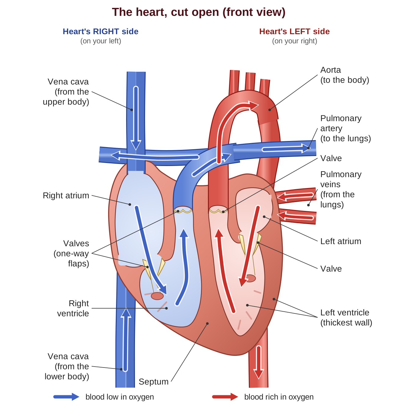
The two sides of the heart carry different kinds of blood. The right side handles deoxygenated blood: blood that has given up most of its oxygen to the body and is on its way to the lungs to pick up more. The left side handles oxygenated blood: blood that has just picked up oxygen in the lungs and is on its way out to the body. The left ventricle has the thickest, strongest wall of all four chambers, because it has the hardest job: it pumps blood out to the entire body, from your head down to your toes.
Here is the path that blood takes through the heart, one step at a time:
- Blood coming back from the body, low in oxygen, enters the right atrium.
- It passes down into the right ventricle, which pumps it out to the lungs.
- In the lungs, the blood drops off carbon dioxide and picks up oxygen.
- The oxygen-rich blood comes back to the heart and enters the left atrium.
- It passes down into the left ventricle, which pumps it out through the body’s largest artery, the aorta, to the whole body.
Two circuits: pulmonary and systemic
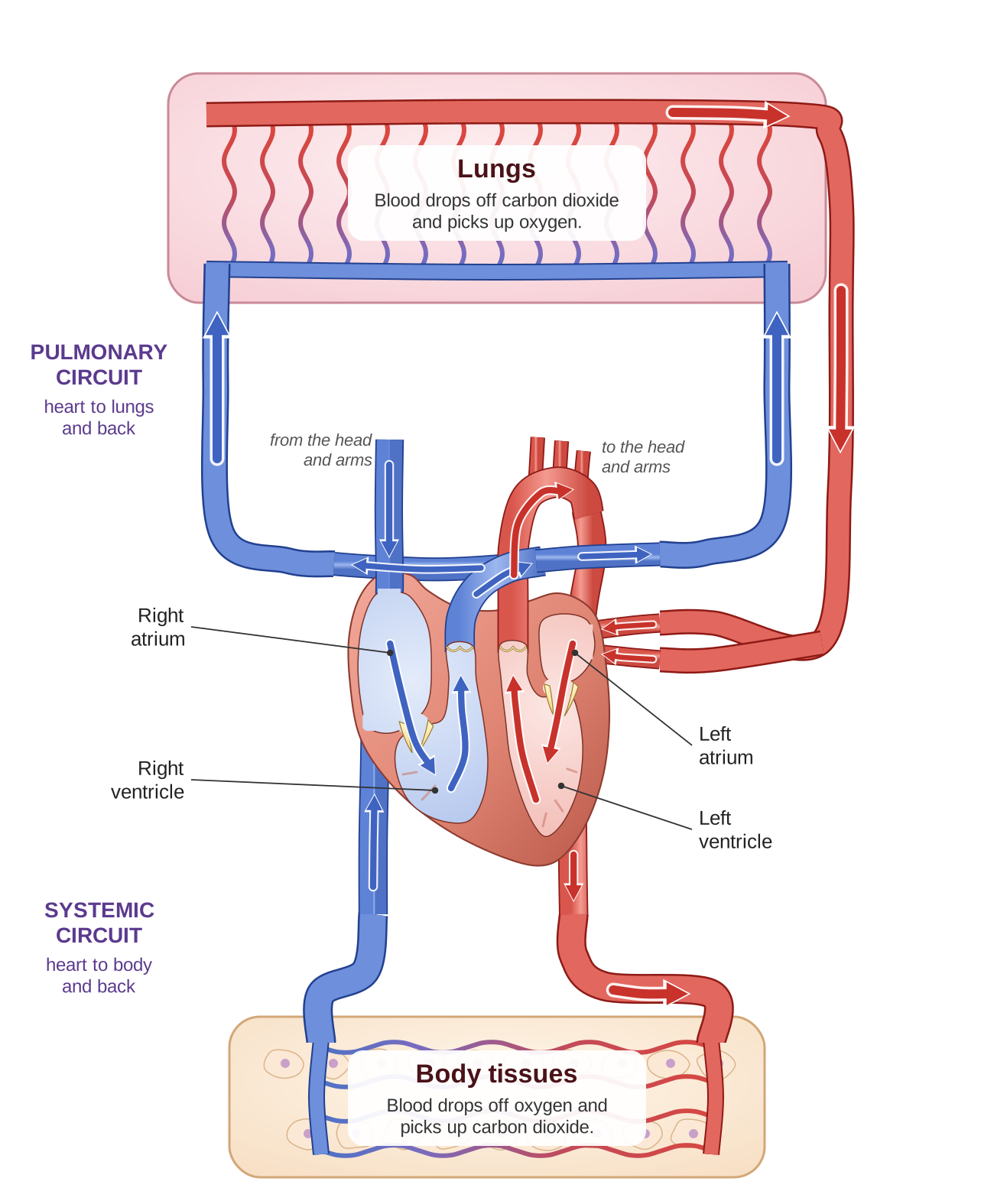
Blood travels in two loops, called circuits. The pulmonary circuit (“pulmonary” means “having to do with the lungs”) carries blood between the heart and the lungs. The systemic circuit carries blood between the heart and the rest of the body.
This means that blood passes through the heart twice on each complete trip: once on its way to the lungs, and once on its way to the body. Scientists call this double circulation.
Blood vessels: the network of tubes
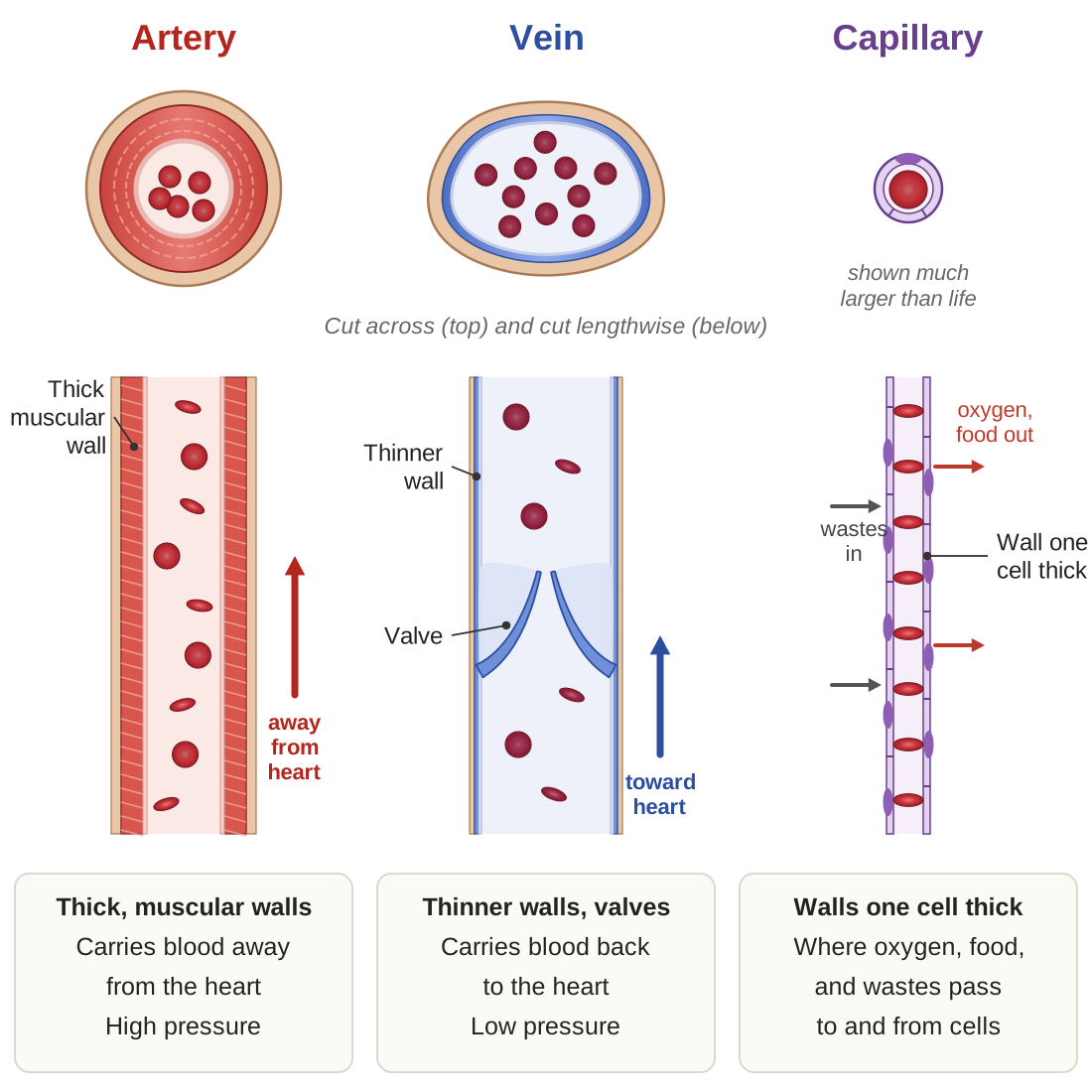
There are three kinds of blood vessels:
| Kind of vessel | What it is like | What it does | Pressure inside |
|---|---|---|---|
| Arteries | Thick, muscular walls | Carry blood away from the heart | High |
| Capillaries | Walls only one cell thick | Where oxygen, food, and wastes pass between blood and cells | Low |
| Veins | Thinner walls, with valves (flaps that keep blood from flowing backward) | Carry blood back to the heart | Low |
“A” is for Artery, and “A” is for Away from the heart.
The respiratory system: getting oxygen in
Air enters through your nose or mouth. It passes down the trachea (the windpipe, the tube in the front of your neck). The trachea splits into two branches called bronchi, one going to each lung. The bronchi keep branching into smaller and smaller tubes, which end in tiny air sacs called alveoli (one is an alveolus). The alveoli are where gas exchange happens. Each alveolus is wrapped in capillaries, and the walls of both the air sac and the capillary are only one cell thick, so gases can pass through them easily.
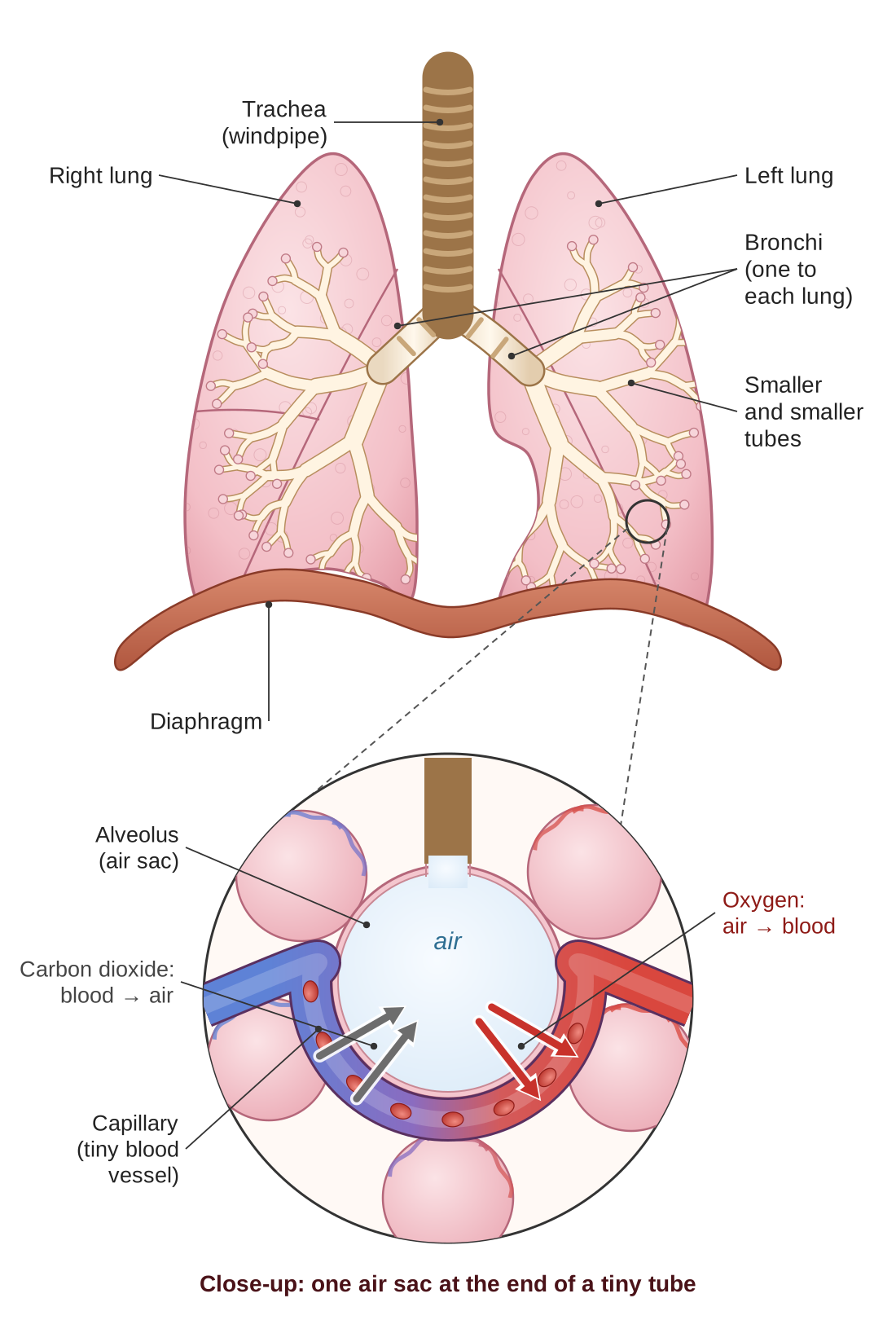
The exchange works by diffusion. Diffusion is the natural movement of a substance from a place where there is a lot of it (a high concentration) to a place where there is less of it (a low concentration). In the lungs:
- Oxygen is plentiful in the air you just breathed in and scarce in the blood arriving from the body, so oxygen moves from the air in the alveolus into the blood.
- Carbon dioxide is plentiful in the blood arriving from the body and scarce in the air in the alveolus, so carbon dioxide moves from the blood into the air in the alveolus. You then breathe it out.
Homeostasis Connection: keeping blood pressure steady
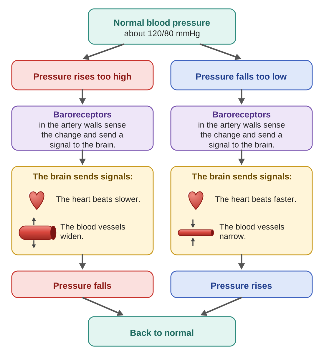
Blood pressure is the force of blood pushing against the walls of the arteries. It is written as two numbers, such as 120/80, and measured in millimeters of mercury (written mmHg), an old unit of pressure. The top number is the pressure while the heart is beating; the bottom number is the pressure while the heart rests between beats. For an adult, a healthy blood pressure is about 120/80 or a little below.
The body keeps blood pressure in a normal range with a negative feedback loop. Sensors called baroreceptors (“baro” means pressure) sit in the walls of certain large arteries. When they detect that pressure has risen or fallen, they send a signal to the brain. The brain then adjusts the heart rate and the width of the blood vessels to bring the pressure back toward normal.
Health Links: when these systems fail
Heart disease. Fatty deposits can build up inside arteries and narrow them, which is often called “clogged” arteries. Narrow arteries make the heart work harder and raise blood pressure. If an artery that feeds the heart muscle itself is blocked, part of the heart is starved of oxygen; this is a heart attack. If an artery that feeds the brain is blocked, part of the brain is starved of oxygen; this is one kind of stroke.
Hypertension is the medical name for blood pressure that stays too high over a long time. Over the years it damages the blood vessels and strains the heart. It is often called the “silent killer,” because most people with high blood pressure feel no symptoms at all until serious damage has been done. That is why it is important to have your blood pressure checked.
Asthma and COPD. In asthma, the airways narrow and swell, often in sudden attacks. COPD (chronic obstructive pulmonary disease) is a long-lasting lung disease, most often caused by smoking. In both, less air moves in and out, so less gas exchange takes place. The person gets too little oxygen and has trouble breathing out carbon dioxide.
Check your understanding
Think it through. What are the four chambers of the heart?
Show a model answer
The right atrium, the right ventricle, the left atrium, and the left ventricle. The two atria are on top and receive blood coming into the heart. The two ventricles are below and pump blood out of the heart.
Think it through. What is the difference between the pulmonary circuit and the systemic circuit?
Show a model answer
The pulmonary circuit carries blood between the heart and the lungs, where the blood drops off carbon dioxide and picks up oxygen. The systemic circuit carries blood between the heart and the rest of the body, where the blood delivers oxygen to the cells and picks up carbon dioxide.
Think it through. How do arteries, capillaries, and veins differ?
Show a model answer
Arteries have thick, muscular walls and carry blood away from the heart under high pressure. Capillaries have walls only one cell thick; they are where oxygen, food molecules, and wastes pass between the blood and the cells. Veins have thinner walls and valves, and they carry blood back to the heart under low pressure.
Think it through. Where does gas exchange happen, and why does it happen there?
Show a model answer
It happens in the alveoli, the tiny air sacs at the ends of the airways in the lungs. It happens there because the walls of the alveoli and of the capillaries around them are only one cell thick, and because there is more oxygen in the air than in the blood, and more carbon dioxide in the blood than in the air. Each gas moves by diffusion from where there is more of it to where there is less.
Think it through. How does the body keep blood pressure steady?
Show a model answer
By negative feedback. Baroreceptors in the arteries sense a change in pressure and signal the brain. If pressure is too high, the brain slows the heart and widens the blood vessels, and pressure falls. If pressure is too low, the brain speeds up the heart and narrows the blood vessels, and pressure rises. In both cases the response undoes the change.
The Digestive System
The digestive system has one main job: to break down the food you eat into molecules small enough to pass into the bloodstream, so that the blood can carry them to your cells. Cells use these molecules for energy, for growth, and for repair.
Digestion happens in two ways. Mechanical digestion is breaking food into smaller pieces physically, by chewing it and by the stomach churning it. Chemical digestion is breaking the large molecules in food into smaller molecules by breaking the chemical bonds that hold them together. Chemical digestion is done by enzymes: proteins that speed up a particular chemical reaction.
The digestive tract: a one-way trip
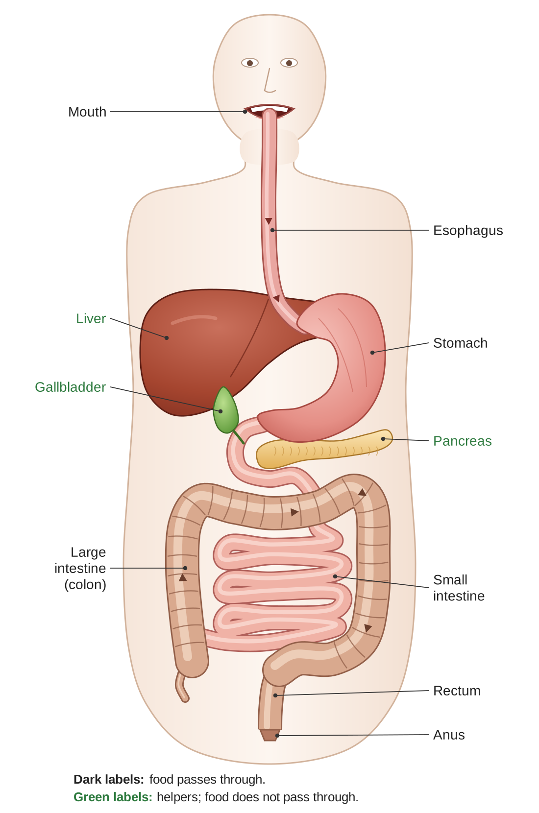
The digestive tract is, in effect, one long tube that runs from the mouth to the anus. It is about 30 feet long, and different stretches of the tube do different jobs. Food moves in only one direction through the tube, pushed along by waves of muscle squeezing called peristalsis. The muscles behind the food squeeze while the muscles ahead of it relax, and this moves the food forward.
The main organs and what they do
Mouth. Digestion begins here. The teeth break food into smaller pieces; this is mechanical digestion. Saliva contains an enzyme called amylase, which begins breaking down starches into sugars; this is chemical digestion.
Esophagus. This is the muscular tube that connects the mouth to the stomach. Peristalsis pushes food down it. Because the muscles do the pushing, and not gravity, you could swallow food even while hanging upside down.
Stomach. The stomach is a muscular bag that churns food while soaking it in hydrochloric acid (a strong acid) and enzymes. The acid kills most bacteria in the food, and it also switches on an enzyme called pepsin, which breaks down proteins. By the time food leaves the stomach, it has become a thick liquid called chyme.
Small intestine. In spite of its name, this is the longest part of the digestive tract, at about 20 feet. (It is called “small” because it is narrow.) It is the main place where nutrients are absorbed, meaning taken into the blood. Its inner wall is covered with millions of tiny finger-shaped bumps called villi. The villi greatly increase the surface area of the wall, that is, the amount of wall that touches the food, so much more absorption can happen at once. Nutrients pass through the wall of the intestine into the bloodstream.
Large intestine (colon). The large intestine takes water and minerals back out of the leftover material. It is home to helpful bacteria, which break down fiber and make certain vitamins. It forms the leftover material into solid waste and stores it until it leaves the body.
The accessory organs: helpers along the way
Several organs help with digestion even though food never passes through them. They are called accessory organs.
Liver. The liver makes bile, a liquid that breaks large drops of fat into many tiny droplets, much as dish soap breaks up grease in a greasy pan. Tiny droplets are easier for enzymes to work on. The liver also processes the nutrients that come to it in the blood from the small intestine.
Gallbladder. This small sac stores bile and makes it stronger, then releases it into the small intestine when it is needed.
Pancreas. The pancreas makes digestive enzymes that break down carbohydrates, proteins, and fats, and sends them into the small intestine. It also makes two hormones (chemical messengers carried in the blood), insulin and glucagon, which control the amount of sugar in the blood. That second job is an important part of homeostasis, and we look at it next.
Homeostasis Connection: keeping blood sugar steady
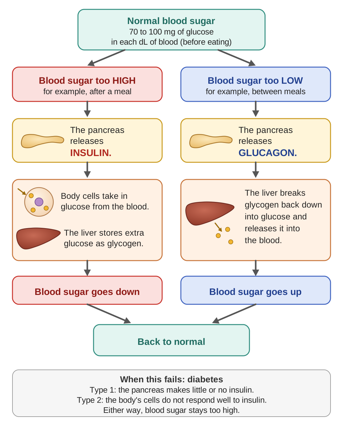
After you eat, carbohydrates in the food are broken down into glucose, a simple sugar, which enters the bloodstream. Cells need the amount of glucose in the blood to stay within a narrow range. Too much or too little causes serious problems. The pancreas keeps this balance with two hormones that have opposite effects:
- Insulin is released when blood sugar is high, for example after a meal. It signals the body’s cells to take glucose in from the blood, and it signals the liver to store extra glucose as glycogen, the form in which animals, including people, store sugar. The result: blood sugar goes down.
- Glucagon is released when blood sugar is low, for example between meals. It signals the liver to break stored glycogen back down into glucose and release it into the blood. The result: blood sugar goes up.
This is a clear example of negative feedback: each hormone works against the change that caused it to be released, and so blood sugar is brought back toward the normal range.
Health Links: when the digestive system fails
Diabetes is the most common disorder of blood sugar control. In Type 1 diabetes, the pancreas makes little or no insulin, so people with Type 1 must take insulin by injection or with a pump. In Type 2 diabetes, the body’s cells stop responding well to insulin’s signal. Insulin is present, but glucose still builds up in the blood. Both types can lead to serious problems if they are not managed.
Obesity. When a person regularly takes in more food energy (calories) than the body uses, the extra energy is stored as fat. Obesity raises the risk of Type 2 diabetes, heart disease, and other conditions.
Malnutrition means not getting enough of the nutrients the body needs. It can come from a poor diet, from digestive disorders that keep the body from absorbing nutrients, or from conditions that raise the body’s need for nutrients.
Digestive disorders. Conditions such as Crohn’s disease, celiac disease, and irritable bowel syndrome affect how well the digestive tract can process food and absorb nutrients.
Check your understanding
Think it through. What is the difference between mechanical digestion and chemical digestion?
Show a model answer
Mechanical digestion breaks food into smaller pieces physically, as when you chew or the stomach churns. Chemical digestion uses enzymes to break the chemical bonds in large food molecules, turning them into smaller molecules. For example, amylase in saliva breaks starch into sugars.
Think it through. Where does most nutrient absorption happen, and why is that organ well suited for the job?
Show a model answer
In the small intestine. It is very long (about 20 feet), and its inner wall is covered with villi, tiny finger-shaped bumps that greatly increase the surface area touching the food. More surface means more nutrients can pass into the blood at once.
Think it through. What do the liver, the gallbladder, and the pancreas each do for digestion?
Show a model answer
The liver makes bile, which breaks fat into tiny droplets, and it processes nutrients coming from the intestine. The gallbladder stores bile and releases it into the small intestine. The pancreas sends digestive enzymes for carbohydrates, proteins, and fats into the small intestine, and it makes insulin and glucagon to control blood sugar.
Think it through. How do insulin and glucagon work together to control blood sugar?
Show a model answer
They have opposite effects. When blood sugar is high, the pancreas releases insulin, which has cells take in glucose and has the liver store glucose as glycogen, so blood sugar falls. When blood sugar is low, the pancreas releases glucagon, which has the liver turn glycogen back into glucose and release it, so blood sugar rises. Together they keep blood sugar within a normal range by negative feedback.
Think it through. What is the difference between Type 1 and Type 2 diabetes?
Show a model answer
In Type 1, the pancreas makes little or no insulin, so the person must take insulin. In Type 2, the pancreas makes insulin, but the body’s cells do not respond to it well, so glucose builds up in the blood anyway.
The Immune System
The immune system is the body’s defense force. It is a network of cells, tissues, and organs that work together to find and destroy invaders from outside the body, while leaving the body’s own cells unharmed. Being able to tell the body’s own cells, called self, apart from anything foreign, called non-self, is at the center of how the immune system works.
Kinds of pathogens
A pathogen is any living thing or agent that can cause disease; people often call pathogens “germs.” The immune system has to defend against several kinds:
| Pathogen | What it is like | Treatment |
|---|---|---|
| Bacteria | Single-celled living things that can reproduce on their own | Antibiotics, which kill bacteria or stop them from growing |
| Viruses | Not cells. Most scientists do not count them as living. A virus can reproduce only by getting inside a living cell and taking over the cell’s machinery to make copies of itself. | Antivirals work against some viruses; vaccines prevent many viral diseases |
| Fungi | Living things whose cells have a nucleus; they include yeasts and molds | Antifungals |
| Parasites | Living things that live on or in a host and feed off it; they range from single-celled organisms to worms | Antiparasitics |
Antibiotics work ONLY against bacteria. They have no effect on viruses. That is why a doctor will not give you antibiotics for a cold or the flu: colds and flu are caused by viruses.
The three lines of defense
The immune system works in layers. If one line of defense fails, the next one takes over.
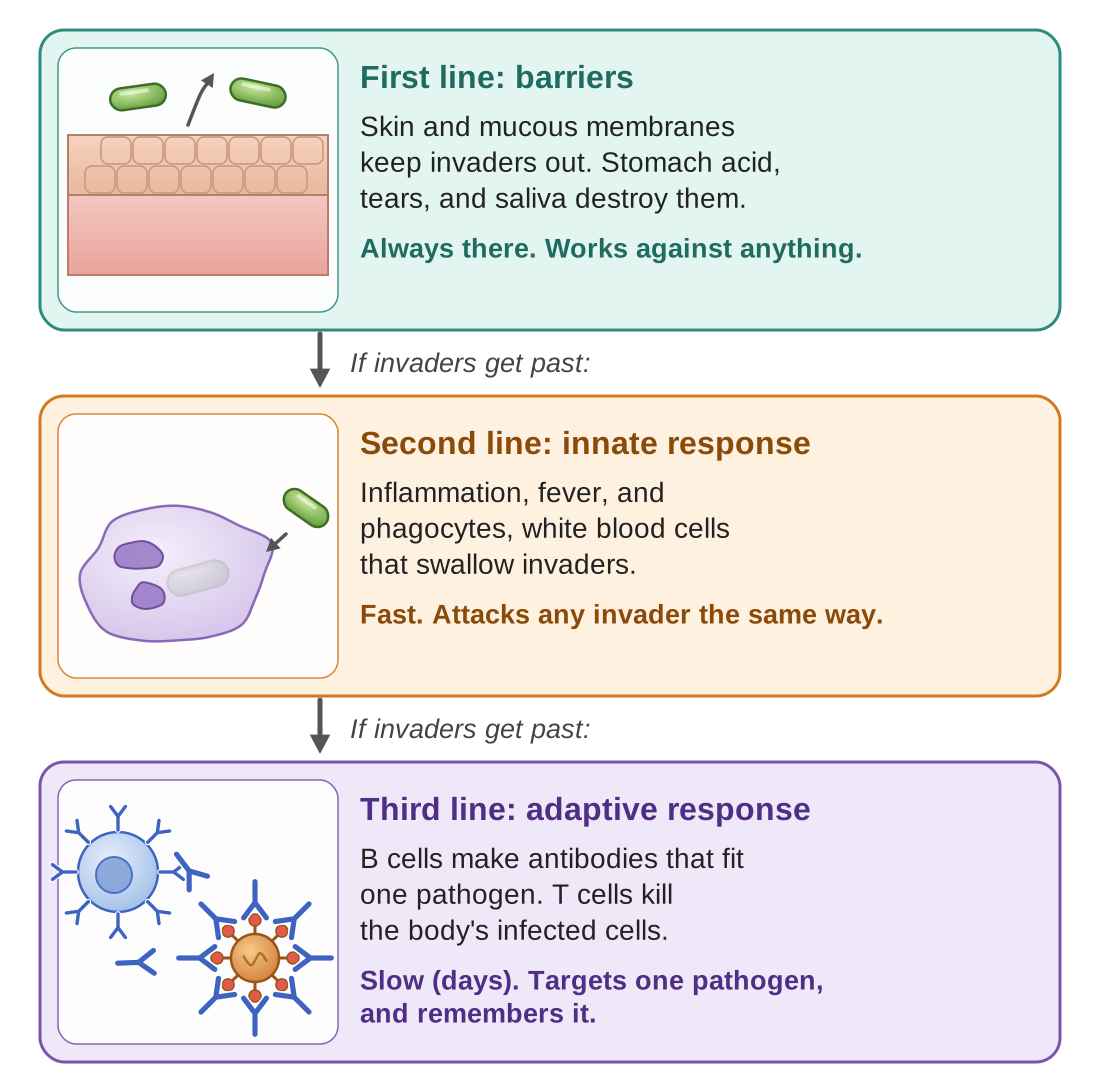
First line: physical barriers. Your skin is the body’s largest organ and its main barrier against pathogens. Mucous membranes, the moist linings of the nose, mouth, lungs, and digestive tract, trap invaders in sticky mucus. Stomach acid kills most pathogens that are swallowed. Tears and saliva contain enzymes that destroy bacteria.
Second line: the innate, or non-specific, response. “Innate” means you are born with it; “non-specific” means it attacks any invader in the same general way. If pathogens get past the first line, the body launches this general counterattack. Inflammation brings extra blood to the area, which is why an inflamed spot is red, warm, and swollen; the extra blood delivers white blood cells. Phagocytes (the name means “eating cells”) are white blood cells that surround invaders, swallow them, and digest them. Fever raises body temperature, which can slow the reproduction of pathogens and speed up the work of immune cells.
Third line: the adaptive, or specific, response. “Adaptive” means it adjusts to each new invader; “specific” means it targets one particular pathogen. If the innate response is not enough, the body mounts a targeted attack. B cells are white blood cells that make antibodies, which mark a particular pathogen for destruction. T cells are white blood cells that kill the body’s own cells once they are infected, or that direct the rest of the immune response. Most importantly, this line creates memory: if the same pathogen returns, the body can respond faster and more strongly than the first time.
Antigens and antibodies: a lock and its key
These two words sound alike, and they are easy to mix up, so take them one at a time. Both come up often on the GED Science test.
An antigen is a molecule, usually a protein, on the surface of a pathogen that the immune system recognizes as foreign. You can think of it as the invader’s identification tag. Each kind of pathogen has its own antigens.
An antibody is a Y-shaped protein made by B cells. The tips of the Y fit onto one particular antigen shape and hold on to it, the way a key fits only one lock. When antibodies attach to a pathogen, they mark it so that other immune cells will destroy it.
So the antigen belongs to the invader, and the antibody is made by your body to match it.
Active and passive immunity
Immunity means protection against a particular disease.
Active immunity develops when your own body makes antibodies, either because you fought off an infection or because you got a vaccine. It takes time to build, but it lasts a long time, often years or a lifetime, because memory cells stay in the body ready to respond.
Passive immunity happens when you receive antibodies that were made somewhere else. Examples are a baby receiving antibodies from its mother through breast milk, or a patient receiving an injection of antibodies. It protects right away, but it does not last, because no memory cells are made.
How vaccines work
Vaccines are among the greatest achievements of medicine. A vaccine trains your immune system to fight a pathogen before you ever meet the real thing.
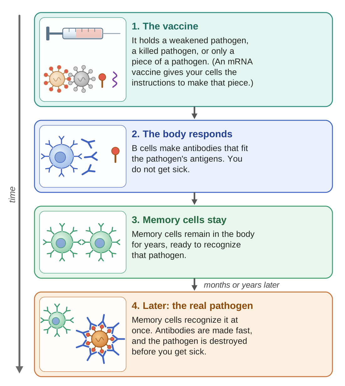
A vaccine contains a weakened pathogen, a killed pathogen, or only a piece of a pathogen. Some newer vaccines, called mRNA vaccines, hold instructions that let your own cells make one harmless piece of the pathogen, so your immune system can learn to recognize it. That is enough to set off an immune response and create memory cells, but not enough to cause the disease. Later, if you meet the real pathogen, your immune system recognizes it right away and destroys it before you get sick.
Homeostasis Connection: telling self from non-self
The immune system must always tell your own cells (self) apart from foreign invaders (non-self). Here is what happens when that fails:
- Autoimmune diseases. The immune system attacks the body’s own tissues by mistake. Examples are Type 1 diabetes (the immune system attacks the cells of the pancreas that make insulin), rheumatoid arthritis (it attacks the joints), and lupus (it attacks many organs).
- Allergies. The immune system overreacts to harmless things, such as pollen, pet dander (tiny flakes of skin from animals), or certain foods, as if they were dangerous pathogens. The inflammation it sets off causes sneezing, itching, and swelling.
- Immunodeficiency. The immune system is weakened and cannot fight infections well. A person can be born with this, or can acquire it later. HIV, the virus that causes AIDS, is an example: it destroys T cells.
Check your understanding
Think it through. What are the four main kinds of pathogens, and why don’t antibiotics work on viruses?
Show a model answer
Bacteria, viruses, fungi, and parasites. Antibiotics work by killing bacteria or stopping their growth, by attacking parts and processes that bacterial cells have. A virus is not a cell and does not have those parts; it reproduces inside the body’s own cells. So antibiotics have nothing in a virus to attack.
Think it through. What are the three lines of immune defense, and how are they different?
Show a model answer
The first line is physical barriers, such as skin, mucous membranes, stomach acid, tears, and saliva, which keep invaders out. The second line is the innate response, such as inflammation, phagocytes, and fever, which acts fast against any invader in the same general way. The third line is the adaptive response, B cells and T cells, which is slower but targets one particular pathogen and remembers it for next time.
Think it through. What is the difference between an antigen and an antibody?
Show a model answer
An antigen is a molecule on the surface of a pathogen, like an identification tag, that the immune system recognizes as foreign. An antibody is a Y-shaped protein made by the body’s B cells that fits one particular antigen, like a key in a lock, and marks the pathogen for destruction.
Think it through. How do vaccines create immunity without causing the disease?
Show a model answer
A vaccine contains a weakened or killed pathogen, or just a piece of one. That is enough for the immune system to recognize its antigens, make antibodies, and create memory cells, but not enough to make you sick. Later, the memory cells recognize the real pathogen and the body destroys it quickly.
Think it through. What happens when the immune system fails to tell self from non-self?
Show a model answer
It can attack the body’s own tissues, which causes autoimmune diseases such as Type 1 diabetes, rheumatoid arthritis, and lupus. A related failure is overreacting to harmless things, which causes allergies.
The Nervous System
The nervous system is the body’s communication and control network. It takes in information from the world around you and from inside your body, makes sense of that information, and directs the body’s responses, often in a fraction of a second. Working alongside the endocrine system (Part E), it maintains homeostasis by adjusting how the body works to meet changing needs.
How it is organized: central and peripheral
The nervous system has two main divisions:
- The central nervous system, or CNS, is the brain and the spinal cord. This is the control center, where information is processed and decisions are made.
- The peripheral nervous system, or PNS, is all the nerves that branch out from the brain and spinal cord to the rest of the body. (“Peripheral” means on the outer edges.) These nerves carry signals to the CNS, such as information from your senses, and from the CNS, such as commands to your muscles.
The neuron: the basic building block
Neurons are nerve cells: cells built to carry electrical signals. They come in many sizes and shapes, but all of them have the same basic parts:
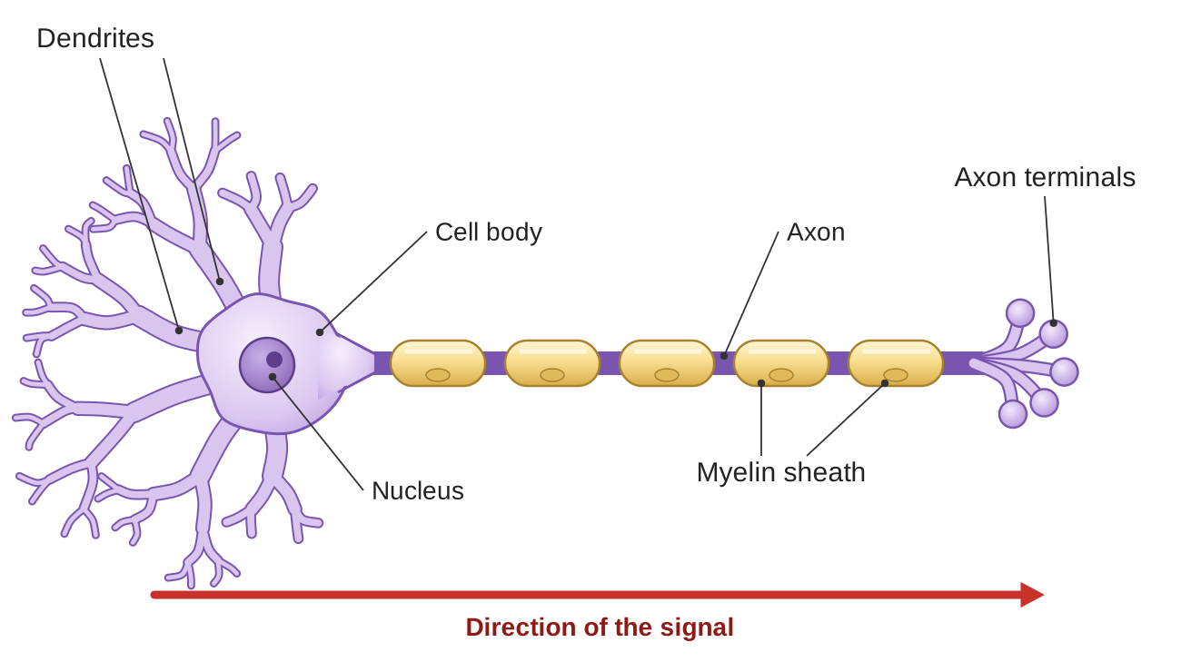
- Dendrites are short branches that receive signals from other neurons. One neuron may have thousands of them.
- The cell body holds the nucleus and adds up the incoming signals. If the signals together are strong enough, the neuron “fires,” that is, it sends a signal of its own.
- The axon is a long fiber that carries the signal away from the cell body toward other neurons or toward the cells the neuron controls, such as muscle cells. Some axons are very long: those that run from the lower spinal cord to the toes can be about three feet long.
- The myelin sheath is a fatty coating that wraps the axon, like the insulation on an electrical wire, and makes the signal travel much faster. The disease multiple sclerosis (MS) damages myelin, which slows nerve signals.
- The axon terminals are the endings of the axon. They release chemical messengers called neurotransmitters into the tiny gap between one neuron and the next. That gap is called a synapse.
How a signal travels: electrical, then chemical
This is a key idea for the GED: a signal travels as an electrical signal inside a neuron, but as a chemical signal between neurons. When the electrical signal reaches the axon terminals, it causes them to release neurotransmitters. These chemicals cross the synapse and attach to receptors on the next neuron. If enough of them attach, the next neuron fires its own electrical signal.
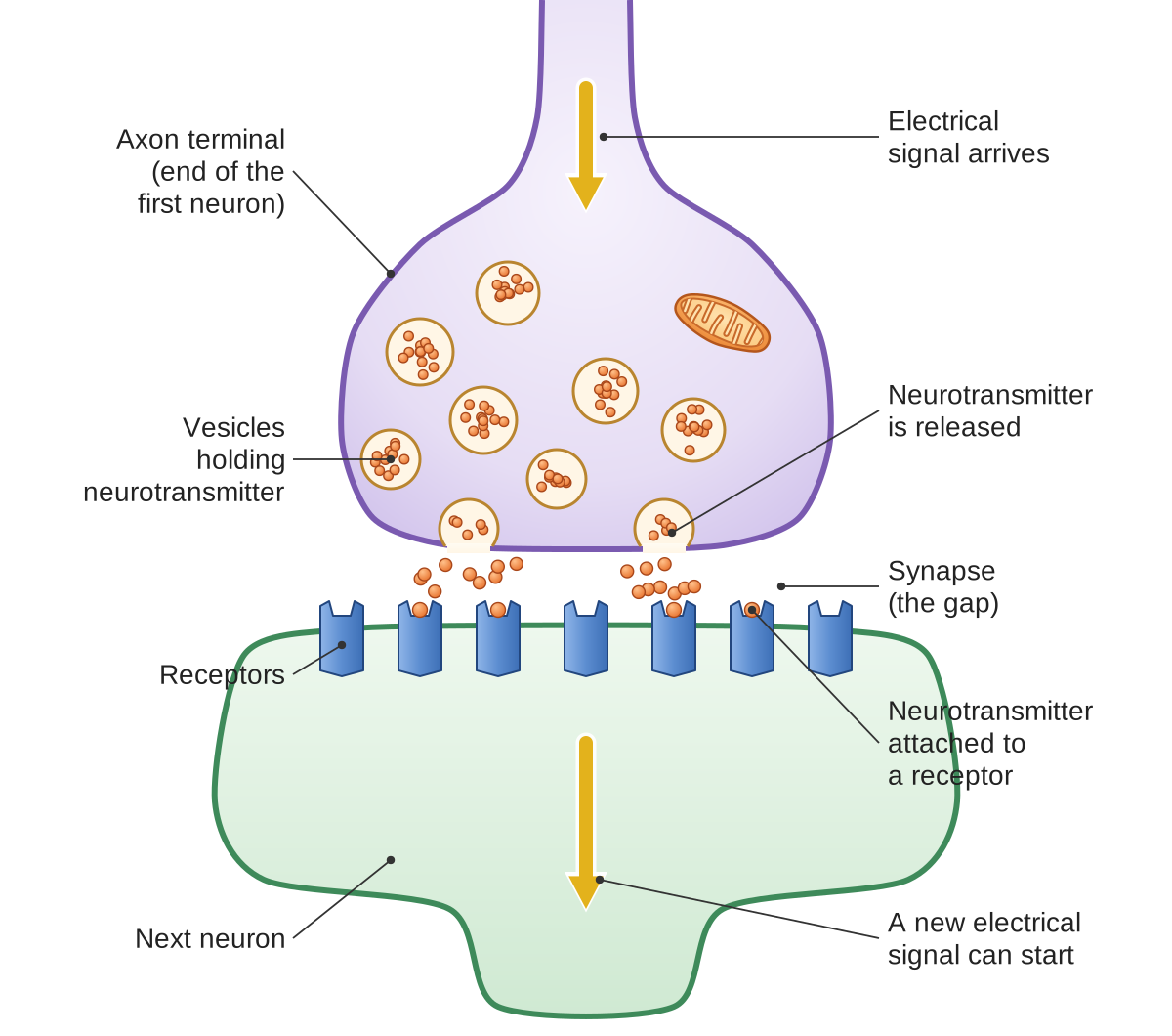
Some common neurotransmitters are dopamine (involved in pleasure and reward), serotonin (involved in mood and sleep), and acetylcholine (which signals muscles to move). Many drugs and medicines work by changing the amount of a neurotransmitter or how strongly it acts.
The reflex arc: responses without thinking
Not every response of the nervous system needs you to think. A reflex is an automatic, very fast response that protects the body from harm. The signal for a reflex follows a set path called a reflex arc:
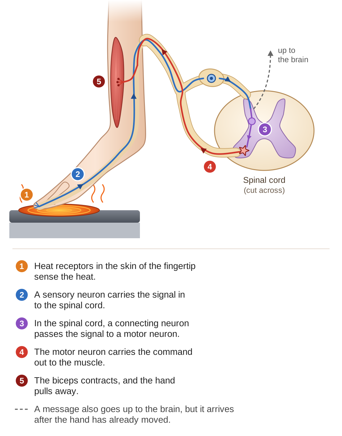
In a reflex, the response comes from the spinal cord and does not wait for the brain. The signal goes in to the spinal cord, and the spinal cord sends the command straight back out to the muscles. Inside the spinal cord, a short connecting neuron passes the signal from the sensory neuron to the motor neuron. A message also travels up to the brain, but it arrives after the reflex is already done. This is why you pull your hand away from a hot stove before you feel the pain.
The brain: the control center
The brain contains about 86 billion neurons, organized into regions that each do particular work. The outer, wrinkled part of the brain is divided into four areas called lobes. For the GED, focus on these regions:
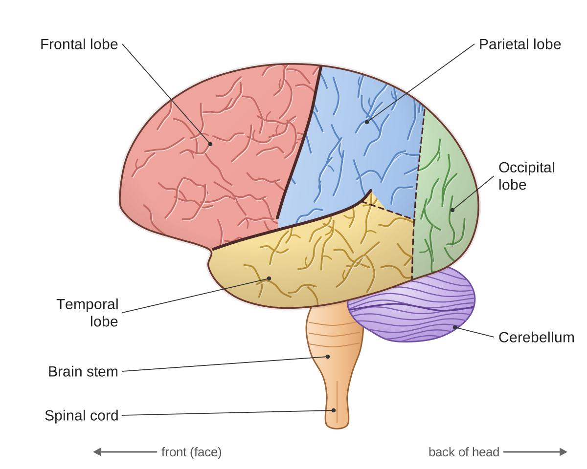
- Frontal lobe (at the front, behind the forehead): personality, decision-making, planning, voluntary movement, and producing speech. Much of what makes you who you are is based here.
- Parietal lobe (at the top, toward the back): handles touch, pressure, temperature, and the sense of where your body is in space.
- Temporal lobe (on each side, near the ears): hearing, forming memories, and understanding language. Damage here can affect a person’s ability to understand speech.
- Occipital lobe (at the back of the head): vision. Visual information is processed mainly here, at the back of the brain, even though your eyes are at the front.
- Cerebellum (below the back of the brain): coordinates movement and balance, and helps you learn movements such as riding a bicycle. Damage causes unsteady, poorly coordinated movement.
- Brain stem (connecting the brain to the spinal cord): controls automatic functions that keep you alive, such as breathing, heart rate, blood pressure, and sleep cycles. Damage here is often fatal.
Homeostasis Connection: “fight or flight” and “rest and digest”
Part of the nervous system, the autonomic nervous system, controls the body functions you do not control on purpose, such as heartbeat and digestion. (“Autonomic” means self-governing, or automatic.) It has two branches:
- The sympathetic branch, called “fight or flight,” switches on during stress or danger. It speeds up the heart rate, widens the pupils of the eyes, sends more blood to the muscles, and signals the adrenal glands to release adrenaline. It gets the body ready for action.
- The parasympathetic branch, called “rest and digest,” switches on during calm times. It slows the heart rate, helps digestion, and saves energy. It returns the body to its normal resting state.
The two branches push the body in opposite directions. Because of this, the body can respond quickly to a change and then return to its normal steady state afterward. In this way they serve the negative feedback that keeps conditions inside the body stable.
Health Links: nervous system disorders
- Alzheimer’s disease. Neurons are lost over time, especially in the areas that control memory. It leads to dementia (a serious loss of memory and thinking ability) and to changes in personality.
- Parkinson’s disease. The neurons that make dopamine are lost. This causes shaking (tremors), stiffness, and difficulty starting a movement.
- Multiple sclerosis (MS). The immune system attacks the myelin sheath. This disrupts nerve signals and causes weakness, numbness, and trouble with coordination.
- Stroke. The blood supply to part of the brain is cut off. Without oxygen, neurons die within minutes. The effects depend on which part of the brain is affected.
Check your understanding
Think it through. What are the two main divisions of the nervous system?
Show a model answer
The central nervous system (CNS), which is the brain and spinal cord, and the peripheral nervous system (PNS), which is all the nerves that branch out from them to the rest of the body.
Think it through. What are the main parts of a neuron, and what does each do?
Show a model answer
Dendrites receive signals from other neurons. The cell body holds the nucleus and decides whether to fire. The axon carries the signal away from the cell body. The myelin sheath insulates the axon and speeds up the signal. The axon terminals release neurotransmitters into the synapse to pass the signal on.
Think it through. How does a signal cross the gap between two neurons?
Show a model answer
When the electrical signal reaches the axon terminals, they release chemical messengers called neurotransmitters into the gap, the synapse. The neurotransmitters cross the gap and attach to receptors on the next neuron, which can start a new electrical signal there.
Think it through. Why are reflexes faster than responses you think about?
Show a model answer
Because the signal only has to travel to the spinal cord and back out to the muscles. The spinal cord sends the command without waiting for the brain, so the path is short. A conscious response needs the signal to travel up to the brain, be processed, and come back down.
Think it through. What do the frontal, temporal, and occipital lobes do?
Show a model answer
The frontal lobe handles personality, decision-making, planning, voluntary movement, and producing speech. The temporal lobe handles hearing, forming memories, and understanding language. The occipital lobe handles vision.
The Endocrine System
The endocrine system is the body’s second communication network. The nervous system uses electrical signals for fast, precise messages. The endocrine system uses hormones: chemical messengers released into the bloodstream, which act more slowly but have effects that last longer. Together, the two systems maintain homeostasis.
Nervous and endocrine: two ways to send a message
Both systems carry information, but they work very differently:
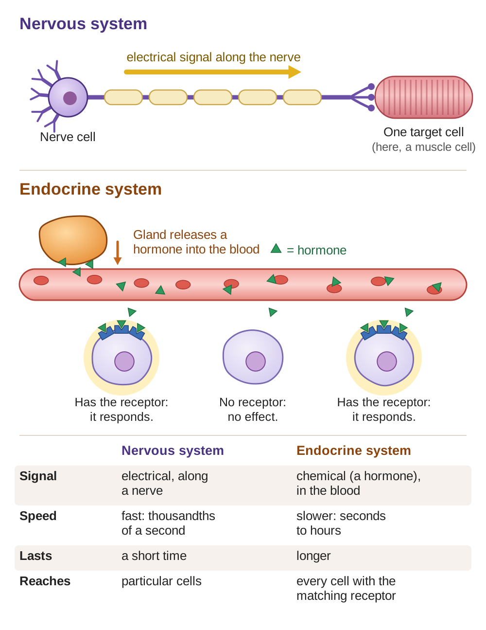
One way to picture the difference: the nervous system is like a telephone call. It connects directly to one person, it is fast, and it ends quickly. The endocrine system is like a letter sent through the mail. It takes longer to arrive, but its message can last longer, and it can reach anyone who has the right mailbox. In the body, the “mailbox” is a receptor, which is explained next.
Hormones: chemical messengers
A hormone is a chemical made by a gland (an organ that makes and releases a substance) and released into the bloodstream. Hormones travel all through the body, but they affect only the cells that have the matching receptor: a protein on or inside the cell that recognizes that one hormone. This is another lock-and-key relationship, like antigens and antibodies.
Because hormones travel in the blood, one gland can affect cells all over the body. This makes hormones well suited to directing changes that happen throughout the body, such as growth, metabolism (all the chemical reactions in the body, including how quickly cells turn food into energy), and reproduction.
The major endocrine glands
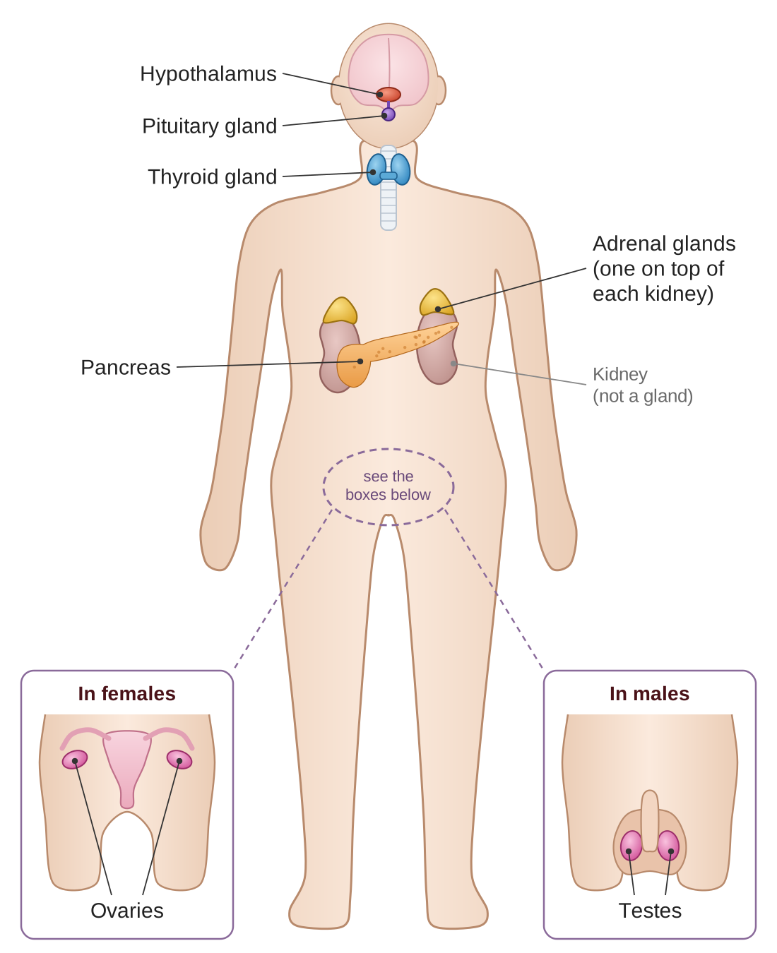
- Hypothalamus. A small area in the brain that links the nervous system and the endocrine system; it is sometimes called the “master regulator.” It controls the pituitary gland, and it keeps track of body conditions such as temperature and hunger.
- Pituitary gland. A pea-sized gland just below the hypothalamus, often called the “master gland” because its hormones control other glands. It makes growth hormone, and it makes hormones that stimulate the thyroid, the adrenal glands, and the reproductive organs.
- Thyroid gland. In the front of the neck. It makes two hormones called T3 and T4 (T4 is also called thyroxine) that control metabolism, that is, how quickly cells turn food into energy.
- Adrenal glands. They sit on top of the kidneys. They make adrenaline, for the fight-or-flight response, and cortisol, for the longer stress response and for control of blood sugar.
- Pancreas. It works both as a digestive organ, making enzymes, and as an endocrine gland, making insulin and glucagon to control blood sugar (see Part B).
- Ovaries and testes. The reproductive glands make the sex hormones: estrogen and progesterone in females, testosterone in males. These hormones control sexual development and reproduction.
Key hormones at a glance
| Hormone | Made by | What it does |
|---|---|---|
| Growth hormone | Pituitary | Stimulates the growth of bones, muscles, and other tissues |
| T3 and T4 | Thyroid | Control metabolism, body temperature, and energy |
| Insulin | Pancreas | Lowers blood sugar (signals cells to take in glucose) |
| Glucagon | Pancreas | Raises blood sugar (signals the liver to release glucose) |
| Adrenaline | Adrenal glands | Fight or flight: speeds the heart and sends more blood to the muscles |
| Cortisol | Adrenal glands | Stress response: raises blood sugar and reduces inflammation |
| Estrogen | Ovaries | Female development; controls the menstrual cycle |
| Testosterone | Testes | Male development, muscle mass, and sperm production |
Hormone feedback loops
Like every system that maintains homeostasis, hormone levels are controlled by negative feedback. When the level of a hormone gets too high, signals cut back its production. When the level drops too low, signals increase its production. The thyroid is a clear example:
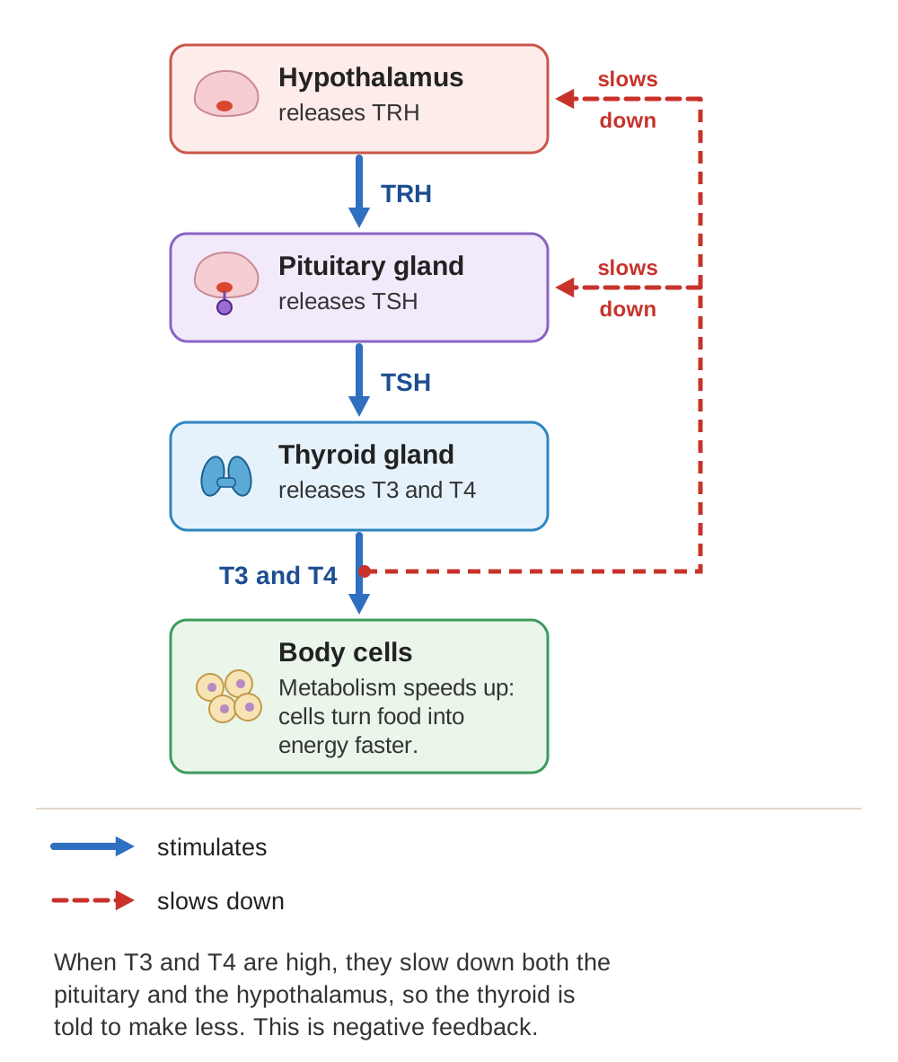
Notice the chain of command: the hypothalamus sends a hormone (TRH) that tells the pituitary to act, and the pituitary sends a hormone (TSH, thyroid-stimulating hormone) that tells the thyroid to act. But the thyroid’s own hormones travel back to both the pituitary and the hypothalamus. When there is enough T3 and T4, they slow both glands down, so the thyroid is told to make less. Control at more than one level keeps the amount very precise.
Homeostasis Connection: the stress response
The endocrine system and the nervous system work together during stress. When you sense danger:
- The nervous system (its sympathetic branch) sets off the immediate fight-or-flight response: a faster heart rate, faster breathing, and sharper alertness.
- The endocrine system releases adrenaline and cortisol, which keep up the stress response: they keep blood sugar raised, keep you alert, and hold back functions that are not urgent, such as digestion.
This is helpful when the threat is short. But when stress goes on for a long time (chronic stress), cortisol stays high, and that can lead to health problems, including a weaker immune system, weight gain, and problems with the heart and blood vessels.
Health Links: endocrine disorders
- Diabetes (explained in Part B) is the most common endocrine disorder. It is a problem with insulin: either not enough of it, or cells that do not respond to it.
- Hypothyroidism means an underactive thyroid that makes too little T3 and T4. (“Hypo” means under.) Symptoms include tiredness, weight gain, feeling cold easily, and depression.
- Hyperthyroidism means an overactive thyroid that makes too much T3 and T4. (“Hyper” means over.) Symptoms include weight loss, a fast heartbeat, anxiety, and feeling too hot easily.
- Growth disorders. Too much growth hormone in childhood causes gigantism, unusually great height. Too little can cause unusually short height; this is one cause of dwarfism. In adults, too much growth hormone causes acromegaly, in which the hands, feet, and face grow larger.
- Adrenal disorders. In Cushing’s syndrome the body has too much cortisol; in Addison’s disease it has too little. Both affect metabolism, the immune system, and the stress response.
Check your understanding
Think it through. How do hormones differ from nerve signals in speed, in how long they last, and in which cells they reach?
Show a model answer
Nerve signals are fast (thousandths of a second), brief, and go along nerves to particular cells. Hormones are slower (seconds to hours), their effects last longer, and they travel in the blood to every cell that has the matching receptor, which can be cells all over the body.
Think it through. What is the chain of command in the endocrine system? (Hint: hypothalamus → ?)
Show a model answer
The hypothalamus controls the pituitary gland, and the pituitary controls other glands, such as the thyroid, the adrenal glands, and the ovaries or testes. Those glands then act on the body’s cells.
Think it through. Which hormones does the pancreas make, and what does each one do?
Show a model answer
Insulin and glucagon. Insulin lowers blood sugar by having cells take in glucose and having the liver store it as glycogen. Glucagon raises blood sugar by having the liver break glycogen down into glucose and release it into the blood.
Think it through. How does negative feedback control the amount of thyroid hormone?
Show a model answer
The hypothalamus releases TRH, which makes the pituitary release TSH, which makes the thyroid release T3 and T4. When T3 and T4 levels get high, they slow down the hypothalamus and the pituitary, so less TSH is sent and the thyroid makes less. When the levels drop, that slowing eases, and production rises again.
Think it through. What are the symptoms of hypothyroidism, and how are they different from those of hyperthyroidism?
Show a model answer
Hypothyroidism (too little thyroid hormone) slows the body down: tiredness, weight gain, feeling cold, and depression. Hyperthyroidism (too much) speeds it up: weight loss, a fast heartbeat, anxiety, and feeling too hot. The symptoms are roughly opposites.
The Muscular System
The muscular system makes movement possible: running and lifting, but also the beating of your heart and the churning of your stomach. Muscles work by contracting, which means shortening. When a muscle contracts, it pulls on whatever it is attached to. Muscles always pull; they never push. Learning the three kinds of muscle, and how contraction works, shows how the build of each kind fits the job it does.
Three kinds of muscle tissue
Your body has three distinct kinds of muscle, each suited to different work:
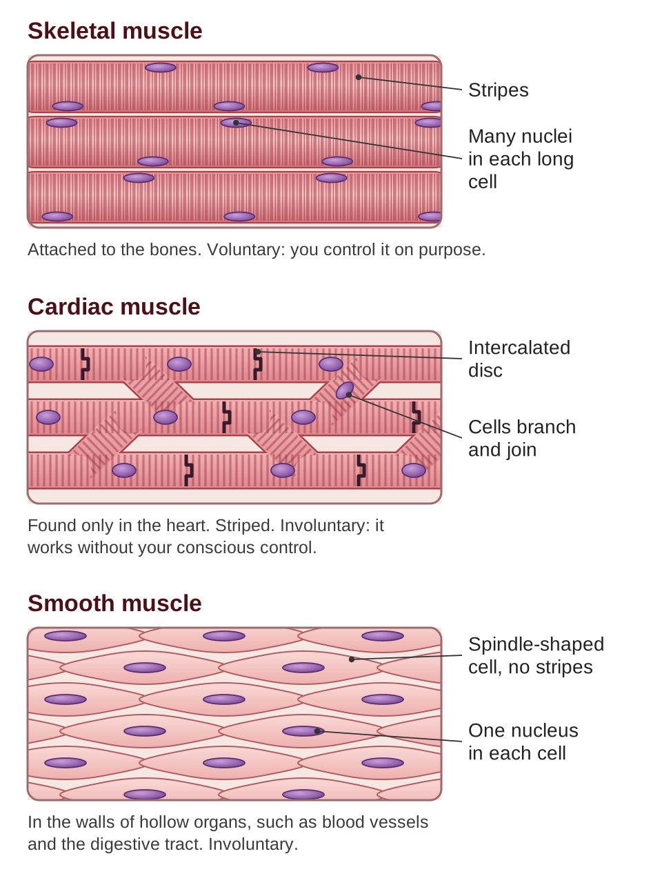
Skeletal muscle is the muscle attached to your bones, the muscle you control on purpose. When you decide to walk, throw a ball, or smile, you use skeletal muscle. Under a microscope it looks striped; the scientific word is striated. The stripes come from the very orderly way its inner parts are arranged. Skeletal muscle cells are unusual: they are very long (some run the whole length of the muscle), and each one has many nuclei.
Cardiac muscle is found only in the heart. It is striped like skeletal muscle, but it works involuntarily, meaning without your conscious control; you do not decide to make your heart beat. Cardiac muscle cells branch and are joined by special connections called intercalated discs. These let the electrical signal spread quickly from cell to cell, so the whole heart contracts together as one unit. Cardiac muscle hardly tires: it contracts about 100,000 times a day without resting.
Smooth muscle is found in the walls of hollow organs: the blood vessels, the digestive tract, the bladder, the airways, and the uterus. It has no stripes, and it too works involuntarily. It contracts slowly, but it can stay contracted for a long time. When your stomach churns food, or your blood vessels narrow to raise blood pressure, smooth muscle is doing the work.
How muscles contract: the sliding filament model
Scientists understand skeletal muscle contraction very well. The key idea is that a muscle does not shorten by squeezing its parts smaller. Instead, two kinds of protein threads, called filaments, slide past each other.
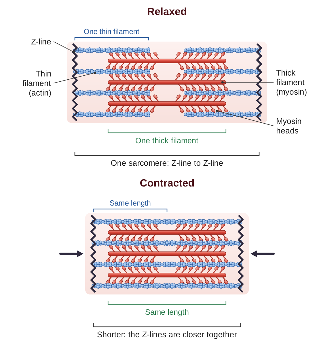
Inside each muscle cell (also called a muscle fiber) are thousands of repeating units called sarcomeres. Each sarcomere is marked off at its two ends by lines called Z-lines. Each sarcomere contains two kinds of protein filaments:
- Thin filaments, made of a protein called actin, are attached to the Z-lines and reach in toward the center of the sarcomere.
- Thick filaments, made of a protein called myosin, sit in the center of the sarcomere. They have small “heads” that can grab onto actin.
During contraction, the myosin heads grab the actin, pull it toward the center, let go, and grab again farther along, much like climbing a rope hand over hand. This pulls the two Z-lines closer together, and the sarcomere gets shorter. When millions of sarcomeres shorten at the same time, the whole muscle contracts.
Energy for contraction
Muscle contraction needs ATP (adenosine triphosphate), the molecule cells use to carry energy for their work. Each cycle of a myosin head grabbing and pulling uses one ATP molecule. Muscles get ATP in three ways:
- Stored ATP and creatine phosphate. Muscles keep a small supply of ready ATP, plus a molecule called creatine phosphate that can quickly make more. Together these give immediate energy for the first few seconds of hard effort.
- Anaerobic respiration (also called lactic acid fermentation). “Anaerobic” means without oxygen. This pathway breaks down glucose without oxygen. It makes ATP quickly but only a little of it, and it produces lactic acid as a by-product. It is good for short, hard bursts, such as a sprint, but muscles working this way tire quickly.
- Aerobic respiration. “Aerobic” means with oxygen. This pathway uses oxygen to break glucose (or fat) down completely, and it makes much more ATP from each glucose molecule. It needs a steady supply of oxygen. It powers long, steady activities such as jogging or cycling.
Muscles work in pairs
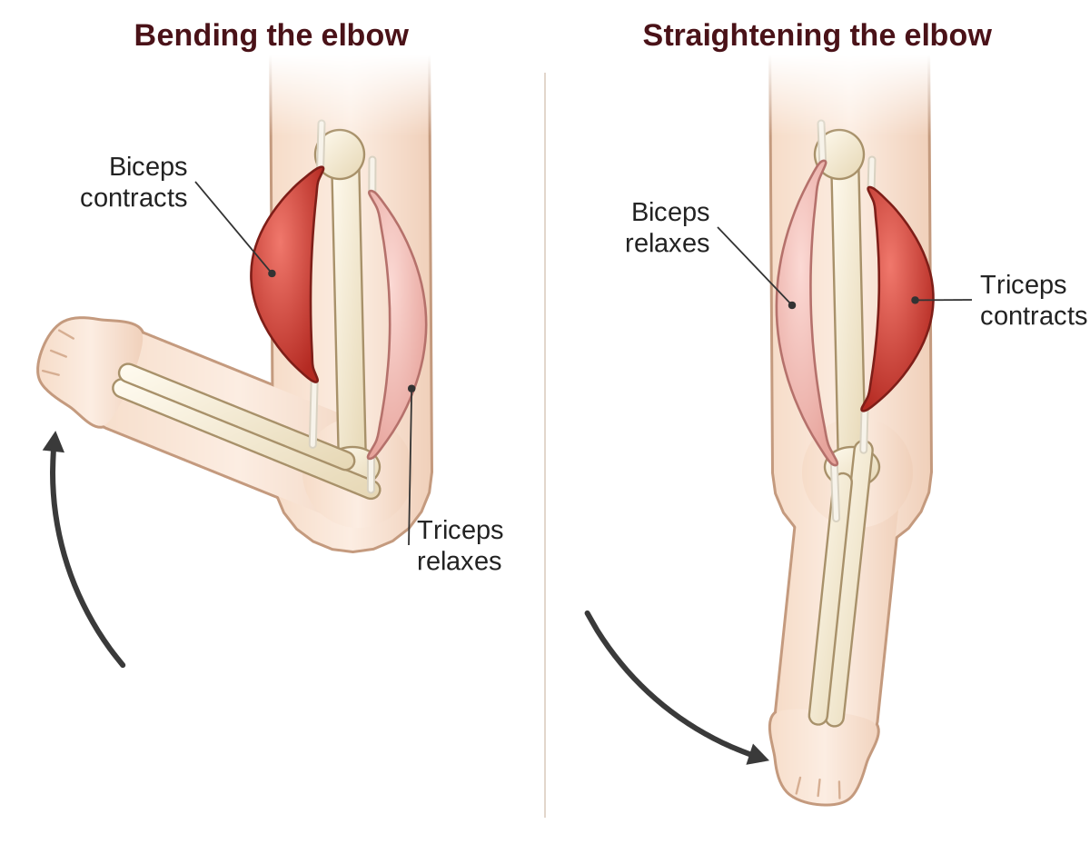
Because muscles can only pull, not push, they usually work in opposing pairs, called antagonistic pairs. When one muscle of the pair contracts, its partner relaxes.
- Biceps and triceps. To bend your elbow, the biceps (on the front of the upper arm) contracts while the triceps (on the back) relaxes. To straighten your elbow, the triceps contracts while the biceps relaxes.
- Quadriceps and hamstrings. These opposing groups of muscles on the front and back of the thigh straighten and bend the knee.
Working in pairs lets the body control movement precisely in both directions.
Homeostasis Connection
The muscular system helps maintain homeostasis in several ways:
- Temperature. Contracting muscles give off heat. Shivering, which is rapid, involuntary contraction of skeletal muscles, is the body’s response to cold: it produces heat to keep the body’s core temperature up.
- Blood pressure. Smooth muscle in the walls of blood vessels narrows or widens the vessels to control blood pressure and to send blood where it is needed.
- Digestion. Smooth muscle contractions (peristalsis) move food through the digestive tract at the right pace.
- Posture. Even when you are standing “still,” your skeletal muscles are partly contracted to hold your posture and keep your balance.
Health Links: muscular system disorders
- Muscular dystrophy is a group of genetic diseases in which the muscles get weaker and break down over time. The most common form, Duchenne muscular dystrophy, is caused by a faulty gene for dystrophin, a protein that helps keep muscle fibers intact.
- Myasthenia gravis is an autoimmune disease. Antibodies block the receptors for acetylcholine at the place where a nerve meets a muscle, so nerve signals cannot make the muscle contract. The result is muscle weakness that gets worse with activity.
- Muscle strains and tears. Stretching or loading a muscle too much can damage its fibers. Minor strains heal with rest; severe tears may need surgery.
- Cramps are sudden, involuntary muscle contractions, often caused by dehydration, an imbalance of electrolytes (minerals such as sodium and potassium dissolved in body fluids), or muscle tiredness.
- Sarcopenia is the loss of muscle mass and strength that comes with age. Regular strength training (exercise against resistance, such as lifting weights) can slow it a great deal.
Check your understanding
Think it through. What are the three kinds of muscle tissue, and where is each found?
Show a model answer
Skeletal muscle is attached to the bones. Cardiac muscle is found only in the heart. Smooth muscle is found in the walls of hollow organs, such as the blood vessels, digestive tract, bladder, airways, and uterus.
Think it through. Which kind of muscle is under voluntary control? Which kinds are involuntary?
Show a model answer
Skeletal muscle is voluntary: you control it on purpose. Cardiac muscle and smooth muscle are involuntary: they work without your conscious control.
Think it through. In the sliding filament model, what actually slides? What provides the energy?
Show a model answer
The thin filaments (actin) slide past the thick filaments (myosin), pulled toward the center of the sarcomere by the myosin heads. The filaments do not get shorter; they overlap more. The energy comes from ATP: each grab-and-pull of a myosin head uses one ATP molecule.
Think it through. Why do muscles work in antagonistic pairs?
Show a model answer
Because a muscle can only pull, not push. To move a joint one way, one muscle pulls; to move it back, a different muscle on the other side has to pull. For example, the biceps bends the elbow and the triceps straightens it.
Think it through. How does the muscular system help control body temperature?
Show a model answer
Contracting muscles give off heat. When the body is cold, skeletal muscles shiver, contracting rapidly and involuntarily, which produces heat and raises body temperature back toward normal.
The Skeletal System
The skeletal system is the framework that supports your body and makes movement possible. But bones are much more than a frame that sits still. They are living tissue: they grow, repair themselves, store minerals, and make blood cells. The adult human skeleton has 206 bones, and the shape of each bone suits its job.
What the skeletal system does
- Support. The skeleton is the rigid frame that holds the body up and gives it its shape. Without bones, your body would have nothing to hold it up and could not keep its shape, somewhat like a jellyfish out of water.
- Protection. Bones shield the most important organs. The skull protects the brain, the rib cage protects the heart and lungs, and the vertebrae (the bones of the spine) protect the spinal cord.
- Movement. Muscles are attached to bones. When the muscles contract, they pull on the bones, and the bones move at the joints.
- Mineral storage. Bones store calcium and phosphorus. About 99 percent of the body’s calcium is in the bones. When the amount of calcium in the blood drops, hormones signal the bones to release some. This is an important part of homeostasis.
- Making blood cells. Red bone marrow, found inside spongy bone, makes red blood cells, white blood cells, and platelets (cell pieces that help blood clot). In adults, this happens mainly in the flat bones (the skull, ribs, pelvis, and breastbone) and in the ends of the long bones.
What bone is made of
Bone is made of living cells set in a hard material, called the matrix, made of calcium phosphate (a mineral) and collagen (a tough, stringy protein). The combination makes bone both strong and slightly flexible. A bone of pure mineral would be brittle and would snap easily; a bone of pure protein would be too soft to hold you up.
A long bone, such as the thigh bone, has these parts:
- Compact bone is the dense, solid outer layer. It gives bone its strength and protection. It looks smooth and solid, but it is actually built of tiny cylinders called osteons, with small channels for blood vessels running through them.
- Spongy bone is full of small open spaces, like a honeycomb. It is found in the ends of long bones and inside flat bones. The open spaces make the bone lighter while keeping it strong, the same way the crisscrossing beams of a bridge are strong without being solid. The spaces hold red marrow, which makes blood cells.
- Bone marrow is the soft tissue inside bones. Red marrow makes blood cells and is found in spongy bone. Yellow marrow stores fat, and in adults it fills the hollow center of the long bones.
- The periosteum is the tough outer skin that covers the surface of bones. It contains nerves (which is why a bone injury hurts), blood vessels that bring nutrients, and cells that are important for bone growth and repair.
Bone cells
Three kinds of cells keep reshaping bone throughout your life:
- Osteoblasts are the builders. They make new bone, putting down the collagen and minerals that form the matrix. When an osteoblast becomes surrounded by the matrix it made, it becomes an osteocyte.
- Osteocytes are the keepers. These mature bone cells keep track of the bone’s condition and respond to the stress put on it. They send signals to each other through tiny channels in the bone to coordinate rebuilding.
- Osteoclasts are the breakers. These large cells break down bone and release its minerals into the blood. That may sound harmful, but it is necessary for reshaping bone, for repairing it, and for keeping blood calcium steady.
Osteoblasts build bone. Osteoclasts carve it away.
Bone remodeling
Bone is always being broken down and rebuilt. This process is called remodeling. It does several things:
- Repair. Tiny damage from everyday activity is repaired all the time.
- Adapting. Bones get stronger in the places where they are put under the most stress. This is why exercise increases bone density (how much bone material is packed into the bone).
- Calcium control. Osteoclasts release calcium into the blood when blood levels drop; osteoblasts put calcium back into bone when there is enough.
In healthy young adults, building and breaking down are in balance. From around age 30, breaking down begins to outpace building, and bone is slowly lost. This loss is faster when a person gets too little calcium or exercise, and, especially in women, when estrogen falls after menopause (the time, usually around age 50, when menstrual periods stop for good).
Joints: where bones meet
A joint (the scientific word is articulation) is a place where two or more bones meet. Different kinds of joints allow different amounts of movement.
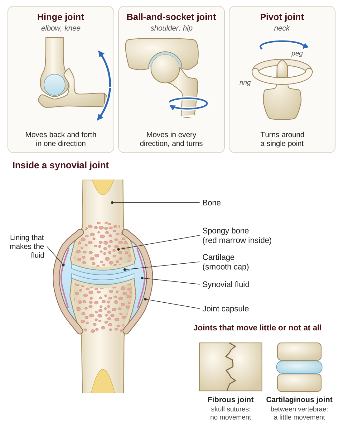
Synovial joints move freely. They contain synovial fluid, a slippery liquid that lets the bones move smoothly. The ends of the bones are capped with smooth cartilage (a firm, rubbery tissue), and the whole joint is enclosed in a capsule. Kinds of synovial joints include:
- Hinge joints (the elbow, the knee) move back and forth in one direction, like a door on its hinge.
- Ball-and-socket joints (the shoulder, the hip) allow movement in every direction, plus turning. They are the most movable kind of joint.
- Pivot joints (in the neck) allow turning around a single point, as when you turn your head to say “no.”
Fibrous joints do not move at all. The bones are held together by dense, tough tissue. The seams between the bones of the skull, called sutures, are fibrous joints.
Cartilaginous joints allow a little movement. The bones are joined by cartilage. Examples are the joints between the vertebrae, and the joint at the front of the pelvis (the pubic symphysis).
The axial and appendicular skeleton
The skeleton is divided into two main parts:
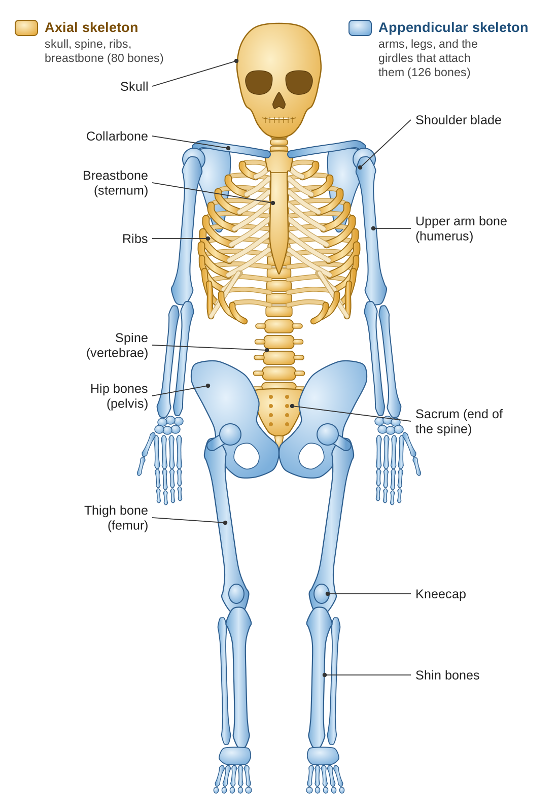
- The axial skeleton (80 bones) runs down the center line of the body: the skull, the spine, the ribs, and the sternum (breastbone). It protects the brain, the spinal cord, and the organs of the chest.
- The appendicular skeleton (126 bones) is the arms and legs, plus the girdles that attach them to the axial skeleton: the shoulder girdle, the arms, and the hands; the hip girdle (pelvis), the legs, and the feet. It is built for movement.
Together, 80 + 126 = 206 bones.
Homeostasis Connection: keeping blood calcium steady
The skeleton plays a central part in keeping the amount of calcium in the blood steady. Two hormones from the endocrine system (Part E) do this work:
- Parathyroid hormone (PTH) comes from the parathyroid glands, four small glands on the back of the thyroid. It is released when blood calcium is low. It stimulates osteoclasts to break down bone, which releases calcium into the blood.
- Calcitonin comes from the thyroid. It is released when blood calcium is high. It holds back the osteoclasts and helps calcium be stored in bone. (In adults, PTH does most of this work; calcitonin plays a smaller part.)
This is another negative feedback loop: a change away from the normal calcium level sets off a response that brings the level back to normal.
Health Links: skeletal system disorders
- Osteoporosis. Bones become porous (full of holes) and fragile because bone is lost: the osteoclasts break down bone faster than the osteoblasts build it. Risk factors include older age, being female, too little calcium or vitamin D, too little exercise, and smoking. It makes broken bones much more likely.
- Arthritis means inflammation of the joints. Osteoarthritis comes from the cartilage in a joint wearing down over time (“wear and tear”). Rheumatoid arthritis is an autoimmune disease in which the immune system attacks the tissues of the joints.
- Fractures are broken bones. In a simple (closed) fracture, the skin is not broken. In a compound (open) fracture, the broken bone comes through the skin. A stress fracture is a tiny crack caused by force repeated over and over. Bones heal by forming a callus, a mass of new tissue that bridges the break and slowly hardens into bone. Healing needs enough calcium, enough vitamin D, and time.
- Rickets and osteomalacia. Bones become soft and weak because of a lack of vitamin D, which the body needs to absorb calcium. Rickets affects children and can cause bowed legs; osteomalacia is the adult form.
Check your understanding
Think it through. What are the five main jobs of the skeletal system?
Show a model answer
Support (holding the body up and giving it shape), protection (of the brain, heart, lungs, and spinal cord), movement (bones are pulled by muscles), mineral storage (especially calcium and phosphorus), and making blood cells (in red marrow).
Think it through. What is the difference between compact bone and spongy bone?
Show a model answer
Compact bone is the dense, solid outer layer that gives bone its strength. Spongy bone has many small open spaces like a honeycomb; it is lighter, it is found in the ends of long bones and inside flat bones, and its spaces hold red marrow, which makes blood cells.
Think it through. What do osteoblasts, osteocytes, and osteoclasts each do?
Show a model answer
Osteoblasts build new bone. Osteocytes are mature bone cells that keep track of the bone’s condition and coordinate rebuilding. Osteoclasts break bone down and release its minerals, such as calcium, into the blood.
Think it through. Name three kinds of synovial joints and give an example of each.
Show a model answer
A hinge joint, such as the elbow or knee. A ball-and-socket joint, such as the shoulder or hip. A pivot joint, such as the joint in the neck that lets you turn your head.
Think it through. How do PTH and calcitonin work together to control the amount of calcium in the blood?
Show a model answer
They have opposite effects. When blood calcium is low, the parathyroid glands release PTH, which has osteoclasts break down bone and release calcium, so blood calcium rises. When blood calcium is high, the thyroid releases calcitonin, which holds back the osteoclasts and helps store calcium in bone, so blood calcium falls. This is negative feedback.
The Integumentary System: Skin, Hair, and Nails
The integumentary system is the skin, the hair, and the nails. (“Integument” means a covering.) The skin is the body’s largest organ. An average adult’s skin covers about 20 square feet and weighs roughly 8 pounds. The skin is not just a wrapping. It is an active organ that protects the body, helps control its temperature, senses the world around it, and even makes vitamin D.
What the integumentary system does
- Protection. Skin is a physical barrier against pathogens, chemicals, and ultraviolet (UV) radiation, the invisible rays in sunlight that can damage cells. Its outer layer is made of dead cells filled with a tough protein called keratin. This layer resists scrapes and keeps the body from losing water. It is your body’s first line of immune defense (see Part C).
- Temperature control. By sweating, by widening or narrowing its blood vessels, and by raising or lowering its hairs, the skin helps keep the body’s core temperature steady. This is an important part of homeostasis.
- Sensation. Skin has millions of sensory receptors that detect touch, pressure, pain, and temperature. They send this information to the nervous system, so that you can respond to what is around you.
- Making vitamin D. When UV light strikes the skin, it starts the first step in making vitamin D, which the body needs to absorb calcium and keep bones healthy.
- Excretion (getting rid of wastes). Sweat glands release small amounts of waste products, such as salts and urea, and help control the body’s water balance.
The layers of the skin
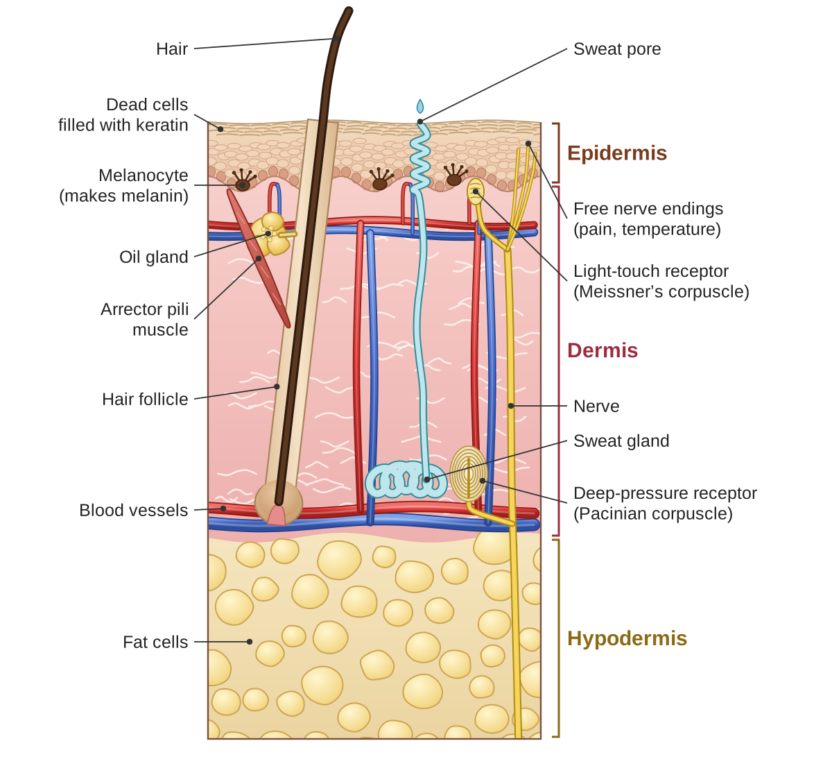
Skin has three layers, and each has its own structures and jobs:
The epidermis
The epidermis is the thin outer layer that you can see and touch. It is made of many stacked layers of cells, and it has no blood vessels. It gets its nutrients by diffusion from the dermis below it.
The epidermis is always renewing itself. Cells at its base divide and are pushed upward. As they rise, they die, fill with keratin, and finally flake off. This cycle takes about two to four weeks. You shed millions of dead skin cells every day.
The main kinds of cells in the epidermis are:
- Keratinocytes, the most common cells, which make keratin for protection and waterproofing.
- Melanocytes, which make melanin, the pigment (coloring substance) that gives skin its color and protects it from UV damage.
- Langerhans cells, immune cells that detect pathogens that get through the skin.
The dermis
The dermis is the thick middle layer, made of connective tissue (tissue that holds other tissues together and supports them). Unlike the epidermis, it has blood vessels and nerves, along with many special structures:
- Hair follicles are the small pockets in the skin that grow hairs. Each follicle has a tiny muscle attached to it, the arrector pili, which contracts to make the hair stand up (goose bumps) when you are cold or frightened.
- Sebaceous glands, or oil glands, release sebum, an oily substance that keeps hair and skin soft and helps kill microbes.
- Sweat glands come in two kinds. Eccrine glands are found all over the body and make watery sweat that cools you. Apocrine glands are found in the armpits and groin and make a thicker sweat; bacteria on the skin break it down, and that causes body odor.
- Sensory receptors are nerve endings of several kinds, each detecting a different sensation: Meissner’s corpuscles detect light touch, Pacinian corpuscles detect deep pressure, and free nerve endings detect pain and temperature.
- Collagen and elastin fibers give the skin strength (collagen) and stretch (elastin). As we age, collagen breaks down and elastin is damaged, and that causes wrinkles.
The hypodermis
The hypodermis, also called the subcutaneous layer (“subcutaneous” means under the skin), lies below the dermis. Strictly speaking it is not part of the skin, but it connects the skin to the tissues underneath. It is made mostly of adipose tissue, which is fat. It keeps the body warm, cushions it, and stores energy.
Homeostasis Connection: temperature control
The skin plays the central role in keeping body temperature at about 37°C (98.6°F):
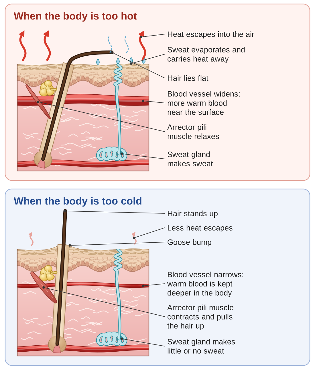
When you are too hot: the blood vessels near the surface of the skin dilate (widen). This brings more warm blood near the surface, where its heat can escape into the air. The sweat glands switch on, and as sweat evaporates (turns from liquid into vapor), it carries heat away from the skin. The hairs lie flat.
When you are too cold: the blood vessels near the surface constrict (narrow), keeping warm blood deeper in the body. The arrector pili muscles contract and raise the hairs, which makes goose bumps. In furry animals, raised fur traps a layer of air that keeps them warm; in humans, who have little body hair, goose bumps do very little, and they are mostly left over from our ancestors. Shivering, which is done by the muscles and not the skin, produces heat.
This is another example of negative feedback: a change away from the normal temperature sets off responses that bring the temperature back to normal.
Skin color
Skin color comes mainly from melanin, the pigment made by melanocytes. People of every skin color have about the same number of melanocytes. The differences in skin color come from how much melanin those cells make, and what kind.
The main job of melanin is to protect the skin from UV radiation. When skin is exposed to sunlight, the melanocytes make more melanin; this is a tan, and it gives some extra protection. But too much UV exposure damages DNA and raises the risk of skin cancer, whatever a person’s skin color.
Other things also affect skin color: carotene, a yellowish pigment that comes from food such as carrots; hemoglobin, the red substance in blood, which gives light skin a pink or reddish tone; and the amount of oxygen in the blood, which is why lips can look blue in the cold.
Hair and nails
Hair is made of dead cells filled with keratin, produced in the hair follicles. Hair gives some protection (on the scalp) and some sensation (you can feel an insect moving on the hairs of your arm). In other mammals it also keeps the body warm. Hair color comes from melanin; hair turns gray as the follicles make less melanin with age.
Nails are also made of cells filled with keratin. They protect the fingertips and help with delicate tasks, such as picking up small objects. The part of the nail you can see is dead tissue; living tissue at the base of the nail, called the nail matrix, makes new nail cells.
Health Links: disorders of the skin
- Skin cancer is the most common kind of cancer. Basal cell carcinoma and squamous cell carcinoma can usually be treated successfully if they are found early. Melanoma, which starts in the melanocytes, is less common but more dangerous. UV exposure is the main risk factor, so sunscreen and staying out of strong sun are the main ways to prevent it.
- Burns are grouped by how deep they go. A first-degree burn affects only the epidermis, as with a sunburn. A second-degree burn reaches into the dermis and causes blisters. A third-degree burn goes through all the layers of the skin and needs skin grafts (skin moved from another part of the body). Severe burns are life-threatening, because the body loses fluid, infection becomes likely, and the skin can no longer control temperature.
- Acne happens when sebum and dead cells clog hair follicles, which often then become infected by bacteria. During puberty, changes in hormones increase the amount of sebum, which is why acne is common in teenagers.
- Eczema and psoriasis are skin conditions involving inflammation. Eczema causes itchy, red, dry patches. Psoriasis causes thick, scaly patches because skin cells are replaced much faster than normal. Both are partly inherited, and both involve the immune system.
- Wound healing. The skin repairs itself in stages: a clot stops the bleeding, inflammation fights infection, new tissue forms, and finally the repair is strengthened and reshaped. A deep wound may leave a scar, where the collagen fibers are laid down in a different pattern from normal skin.
Check your understanding
Think it through. What are the five main jobs of the integumentary system?
Show a model answer
Protection (a barrier against pathogens, chemicals, UV radiation, and water loss), temperature control, sensation (touch, pressure, pain, temperature), making vitamin D, and excretion of small amounts of waste in sweat.
Think it through. What are the three layers of skin, and what does each contain?
Show a model answer
The epidermis, the thin outer layer, has no blood vessels and contains keratinocytes, melanocytes, and Langerhans cells. The dermis, the thick middle layer, contains blood vessels, nerves, hair follicles, oil glands, sweat glands, sensory receptors, and collagen and elastin fibers. The hypodermis, below, is mostly fat tissue that insulates, cushions, and stores energy.
Think it through. How does the skin help control body temperature when you are too hot, and when you are too cold?
Show a model answer
When you are too hot, blood vessels near the surface widen to let heat escape, and sweat glands release sweat, which carries heat away as it evaporates. When you are too cold, blood vessels near the surface narrow to keep warm blood deeper inside, and the hairs rise (goose bumps); the muscles also shiver to make heat.
Think it through. What produces skin color, and what is melanin’s main job?
Show a model answer
Skin color comes mainly from melanin, made by melanocytes; differences in color come from how much melanin is made and what kind, not from the number of melanocytes. Melanin’s main job is to protect the skin from UV radiation.
Think it through. What are the three main kinds of skin cancer, and what is the main risk factor?
Show a model answer
Basal cell carcinoma, squamous cell carcinoma, and melanoma, which is the most dangerous. The main risk factor is exposure to UV radiation, mostly from the sun.
The Urinary (Excretory) System
The urinary system, also called the excretory system, removes wastes from the blood and controls how much water and how many electrolytes (dissolved minerals, such as sodium and potassium) the body holds. People often think of the kidneys as simple filters, but they do much more: they play a central part in homeostasis, keeping the fluid around the cells exactly the way the cells need it to be.
What the urinary system does
- Excretion (getting rid of wastes). The kidneys remove the wastes made by the body’s chemical reactions, especially urea (left over from breaking down proteins), creatinine (left over from muscle activity), and extra ions. Without this, these wastes would build up and poison the body.
- Water balance. The kidneys control the amount of water in the body by changing how concentrated the urine is. When you are dehydrated, they save water by making a small amount of dark, concentrated urine. When you have taken in extra water, they make a lot of pale, watery urine.
- Electrolyte balance. The kidneys keep the right levels of sodium, potassium, calcium, and other electrolytes in the blood. These are needed for nerves and muscles to work.
- pH balance. pH is a measure of how acidic or basic a liquid is. Blood must stay within a narrow pH range, 7.35 to 7.45, for the body’s enzymes to work. The kidneys help keep it there by getting rid of hydrogen ions (which make blood more acidic) or holding on to them.
- Blood pressure control. The kidneys affect blood pressure by holding on to water or letting it go, and by releasing an enzyme called renin, which starts a chain of hormone signals that raises blood pressure.
The parts of the urinary system
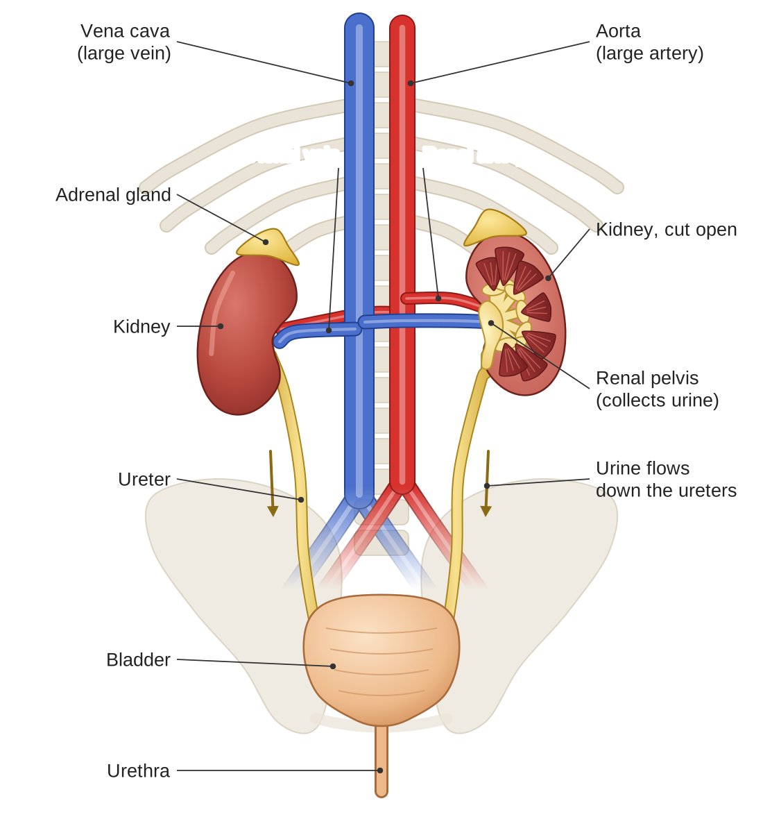
- Kidneys. Two bean-shaped organs at the back of the abdomen, just below the rib cage. Each kidney receives blood through a renal artery and sends the cleaned blood back through a renal vein. (“Renal” means having to do with the kidneys.) About 1,700 liters of blood pass through the kidneys each day. From that blood, the kidneys filter out about 180 liters of fluid, return almost all of it to the blood, and make about 1 to 2 liters of urine.
- Ureters. Two muscular tubes that carry urine from the kidneys down to the bladder. They move it by peristalsis, the same waves of muscle squeezing that move food through the digestive tract.
- Urinary bladder. A hollow, muscular bag that stores urine until it leaves the body. It can comfortably hold about 400 to 600 milliliters (roughly 1.5 to 2.5 cups), though you usually begin to feel the urge to urinate at around 200 milliliters.
- Urethra. The tube through which urine leaves the body. It is much shorter in women (about 4 centimeters, or 1.5 inches) than in men (about 20 centimeters, or 8 inches). This is one reason urinary tract infections are more common in women: bacteria have a shorter distance to travel.
The nephron: the kidney’s working unit
Each kidney contains about one million tiny filtering units called nephrons. To understand how the kidneys work, you need to understand the nephron.
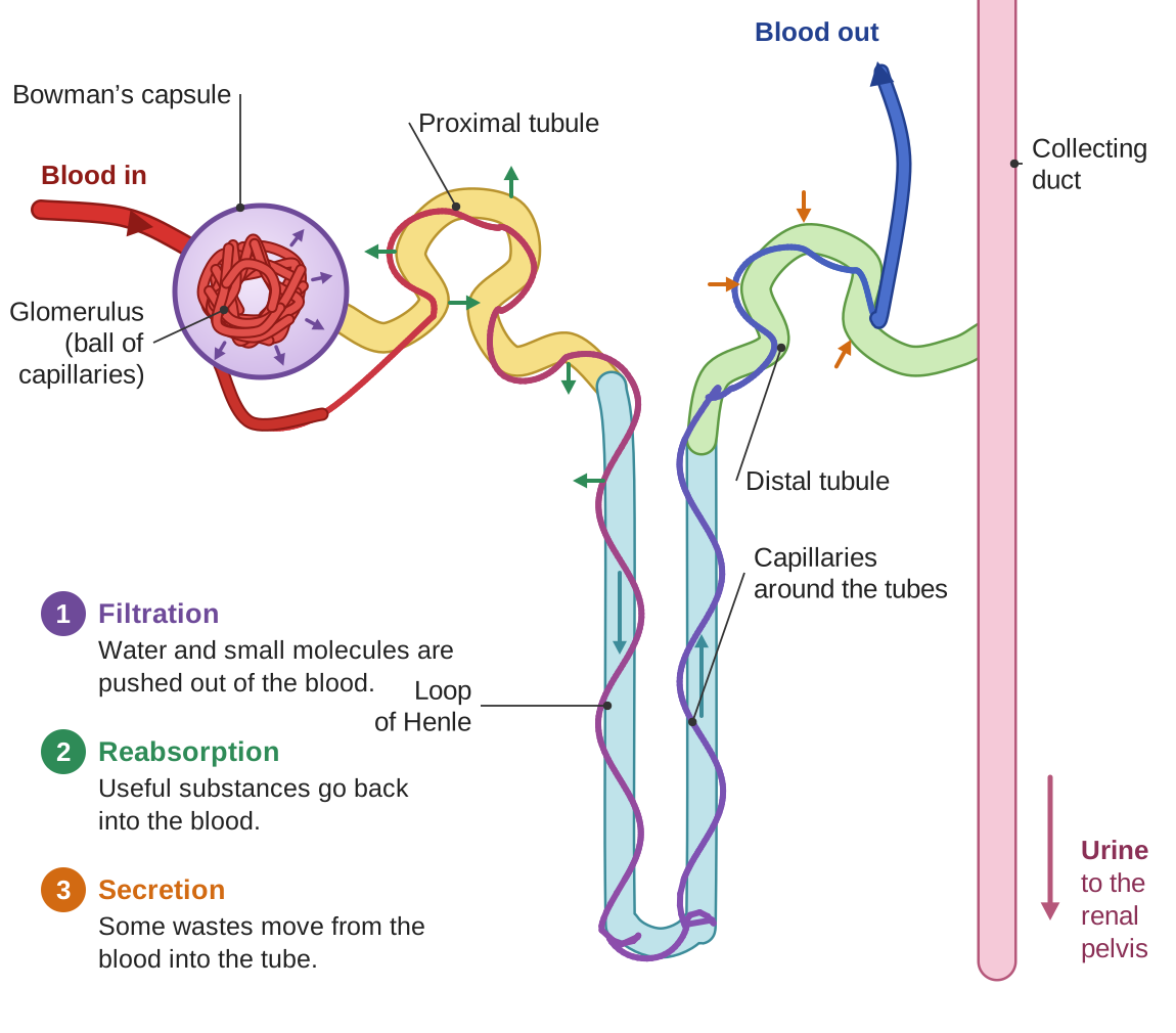
The three steps in making urine
- Filtration. Blood enters the glomerulus, a ball of capillaries, under high pressure. The pressure forces water and small molecules (glucose, amino acids, salts, and urea) out through the capillary walls into a cup around the glomerulus called Bowman’s capsule. The fluid that is filtered out is called filtrate. Large molecules, such as proteins, and blood cells are too big to pass through, so they stay in the blood. About 180 liters of filtrate are made each day, but most of it is taken back, as the next step explains.
- Reabsorption. As the filtrate flows along the tubes of the nephron, useful substances are taken back into the blood through the capillaries that wrap around the tubes. The proximal tubule (the first stretch of tube) takes back about 65 percent of the filtered water, all of the glucose and amino acids (in a healthy person), and much of the salt. The loop of Henle, the U-shaped section, sets up conditions that let the kidney make concentrated urine. The distal tubule (the later stretch) makes fine adjustments to salt and water balance, under the control of hormones.
- Secretion. Some substances are moved from the blood into the tube, to be removed in the urine. These include hydrogen ions (for pH control), potassium, and certain drugs and poisons. This lets the kidneys get rid of substances that were not filtered out, or that need extra removal.
The final urine, which is only about 1 percent of the original filtrate, collects in the collecting duct. From there it drains into a funnel-shaped space in the kidney (the renal pelvis), and then into the ureter.
Homeostasis Connection: water balance
The kidneys work closely with the endocrine system to keep the body’s water in balance:
- Antidiuretic hormone (ADH) is released by the pituitary gland when the blood becomes too concentrated, as it does when you are dehydrated. (“Antidiuretic” means “against making a lot of urine.”) ADH makes the walls of the collecting ducts let more water through, so more water is taken back into the blood. The result is a small amount of concentrated urine. When you are well hydrated, the amount of ADH falls, and the kidneys make larger amounts of watery urine.
- Aldosterone is released by the adrenal glands when blood pressure or blood sodium is low. It makes the distal tubule take back more sodium. Water follows the sodium back into the blood, which increases the volume of blood and raises blood pressure.
This is negative feedback at work: when the concentration of the blood or the blood pressure moves away from normal, hormones set off responses that bring it back.
What is in urine?
Normal urine is about 95 percent water and 5 percent dissolved substances:
- Urea, the largest part of the dissolved material; it is waste from breaking down proteins.
- Creatinine, waste from muscle activity.
- Uric acid, waste from breaking down nucleic acids (the family of molecules that includes DNA).
- Ions such as sodium, potassium, and chloride, in amounts that vary.
- Small amounts of hormones, vitamins, and other molecules.
Normal urine should not contain glucose (glucose in urine suggests diabetes), protein (which suggests kidney damage), or blood (which suggests an injury or disease).
Health Links: urinary system disorders
- Kidney stones are hard lumps of minerals and salts that form in the kidneys. Small stones may pass out through the ureter, often painfully; large ones may need medical treatment. Risk factors include dehydration, a diet high in salt, and certain medical conditions.
- Urinary tract infections (UTIs) are usually caused by bacteria entering the urethra. They are more common in women because of the shorter urethra. Symptoms include pain when urinating, a frequent urge to urinate, and cloudy urine. Most are easily treated with antibiotics, but they can become serious if they spread to the kidneys.
- Chronic kidney disease (CKD) is a gradual loss of kidney function over months or years. (“Chronic” means lasting a long time.) Common causes are diabetes, because high blood sugar damages the nephrons, and high blood pressure, because high pressure damages the glomeruli. In advanced stages, a person may need dialysis or a kidney transplant.
- Kidney failure (also called renal failure) means the kidneys can no longer filter the blood well enough. Acute kidney failure (sudden failure) can come from a severe injury, an infection, or a drug that harms the kidneys, and it may be reversible. Chronic kidney failure gets worse over time and may require dialysis: treatment with a machine that filters the blood when the kidneys cannot.
- Incontinence is loss of bladder control. It can result from weak muscles, nerve damage, prostate problems in men, or other causes. It can often be treated with exercises, medicine, or surgery.
Check your understanding
Think it through. What are the five main jobs of the urinary system?
Show a model answer
Excretion of wastes such as urea and creatinine; water balance; electrolyte balance; pH balance of the blood; and helping to control blood pressure.
Think it through. What are the four main organs of the urinary system, and what does each do?
Show a model answer
The kidneys filter the blood and make urine. The ureters carry urine from the kidneys to the bladder. The bladder stores urine. The urethra carries urine out of the body.
Think it through. What are the three steps in making urine, and where does each happen?
Show a model answer
Filtration happens where the glomerulus meets Bowman’s capsule: pressure pushes water and small molecules out of the blood. Reabsorption happens along the tubules (the proximal tubule, the loop of Henle, and the distal tubule): useful substances such as glucose, amino acids, salt, and most of the water go back into the blood. Secretion also happens along the tubules: substances such as hydrogen ions, potassium, and some drugs are moved from the blood into the tube.
Think it through. How does ADH help keep the body’s water in balance?
Show a model answer
When the blood is too concentrated, as in dehydration, the pituitary releases ADH. ADH makes the collecting ducts let more water back into the blood, so the body keeps water and makes a small amount of concentrated urine. When you are well hydrated, less ADH is released, and the kidneys let more water go as dilute urine.
Think it through. Which substances should NOT normally be found in urine, and what might their presence mean?
Show a model answer
Glucose, which may mean diabetes; protein, which may mean kidney damage; and blood, which may mean an injury or a disease.
The Reproductive System
The reproductive system is different from all the other body systems. It is the only one that a person does not need in order to stay alive, yet the human species needs it in order to continue. This system makes the sex cells, called gametes, makes fertilization possible, and in females supports the growth of a new baby. It also makes hormones that drive sexual development and affect many other body processes.
What the reproductive system does
- Making gametes. The reproductive system makes the sex cells: sperm in males and eggs (also called ova) in females. These are the only cells made by meiosis, the kind of cell division that produces sex cells. Each gamete has one set of 23 chromosomes, instead of the two sets (46 chromosomes) in the body’s other cells. When a sperm and an egg join, the new cell has two sets again: 46.
- Making hormones. The reproductive organs make the sex hormones, which drive puberty (the years when a child’s body matures into an adult’s), keep the reproductive system working, and affect many other body systems. The key reproductive hormones are testosterone (mainly in males) and estrogen and progesterone (mainly in females).
- Fertilization and pregnancy. The system makes it possible for a sperm and an egg to join, and in females it provides a place for the developing baby to grow.
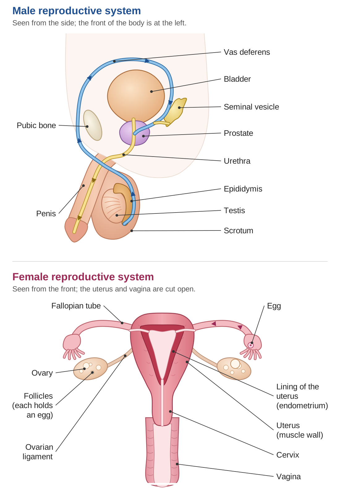
The male reproductive system
- Testes (one is a testis) are the main male reproductive organs, also called the male gonads (the organs that make gametes). They hang in the scrotum, a pouch of skin outside the body, because sperm production needs a temperature slightly lower than normal body temperature. The testes make sperm and testosterone.
- The epididymis is a coiled tube on the surface of each testis, where sperm mature and are stored.
- The vas deferens is a muscular tube, one from each side, that carries sperm from the epididymis toward the urethra during ejaculation.
- The seminal vesicles and the prostate are glands that make the fluid parts of semen (the fluid that carries sperm). The seminal vesicles add fructose, a sugar that gives the sperm energy. The prostate adds enzymes and substances that protect the sperm from acid.
- The penis delivers sperm into the female reproductive tract. The urethra runs through it and carries both urine and semen, though never at the same time.
The female reproductive system
- Ovaries are the main female reproductive organs, the female gonads. Each ovary holds thousands of follicles, small sacs that each hold an immature egg. The ovaries make eggs and the hormones estrogen and progesterone.
- The fallopian tubes (also called oviducts) carry eggs from the ovaries toward the uterus. Fertilization usually happens here.
- The uterus (womb) is a muscular organ where a fertilized egg attaches and grows during pregnancy. Its inner lining, the endometrium, thickens every month to prepare for a possible pregnancy.
- The cervix is the narrow lower end of the uterus, which opens into the vagina. It widens during childbirth.
- The vagina is the birth canal, and the place where sperm are deposited during intercourse.
The menstrual cycle
The menstrual cycle is a monthly cycle, about 28 days long on average, that prepares the female body for a possible pregnancy. It is controlled by hormones from the hypothalamus, the pituitary gland, and the ovaries. Two of the pituitary hormones are FSH (follicle-stimulating hormone) and LH (luteinizing hormone).
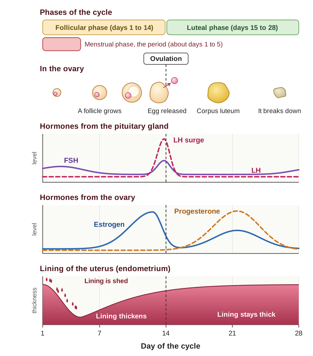
- Menstrual phase (about days 1 to 5). If no pregnancy happened in the last cycle, hormone levels have dropped, and the lining of the uterus is shed. This is menstruation, the monthly period.
- Follicular phase (days 1 to 14). FSH stimulates follicles in the ovary to develop. The growing follicles make estrogen, which makes the lining of the uterus thicken again.
- Ovulation (about day 14). A sudden rise, or surge, in LH triggers ovulation: the release of a mature egg from the ovary. The days just before ovulation and the day of ovulation are when pregnancy is most likely.
- Luteal phase (days 15 to 28). The empty follicle that released the egg becomes a structure called the corpus luteum, which makes progesterone. Progesterone keeps the lining of the uterus ready. If no pregnancy happens, the corpus luteum breaks down, hormone levels fall, and menstruation begins again.
Fertilization and early development
If a sperm reaches an egg in the fallopian tube, fertilization may happen: the sperm and egg join into one cell, called a zygote. The zygote begins dividing as it travels down to the uterus. By the time it attaches to the wall of the uterus (this is called implantation), it has become a hollow ball of cells called a blastocyst. The developing baby is called an embryo for the first 8 weeks, and a fetus from then until birth.
The reproductive hormones
- Testosterone is the main male sex hormone. It drives male development and keeps sperm production going.
- Estrogen is the main female sex hormone. It drives female development and controls the menstrual cycle.
- Progesterone prepares the uterus for pregnancy and keeps it ready.
- FSH and LH are hormones from the pituitary gland that control the making of gametes and the release of sex hormones, in both men and women.
Health Links: reproductive system problems
- Infertility means being unable to get pregnant after a year of trying. Causes include hormone imbalances, problems in the structure of the reproductive organs, and a low sperm count.
- Sexually transmitted infections (STIs) are infections spread through sexual contact. Examples include chlamydia, gonorrhea, HIV, and HPV (human papillomavirus).
- Endometriosis is a condition in which tissue like the lining of the uterus grows outside the uterus. It causes pain and sometimes infertility.
- PCOS (polycystic ovary syndrome) is a hormone disorder that causes irregular periods and often many small fluid-filled sacs (cysts) on the ovaries.
Check your understanding
Think it through. What are the three main jobs of the reproductive system?
Show a model answer
Making gametes (sperm and eggs), making sex hormones, and making fertilization and pregnancy possible.
Think it through. What do the testes make? What do the ovaries make?
Show a model answer
The testes make sperm and testosterone. The ovaries make eggs and the hormones estrogen and progesterone.
Think it through. What are the four phases of the menstrual cycle?
Show a model answer
The menstrual phase (the lining is shed), the follicular phase (follicles develop and the lining thickens), ovulation (an egg is released), and the luteal phase (the corpus luteum makes progesterone and keeps the lining ready).
Think it through. Where does fertilization usually happen?
Show a model answer
In a fallopian tube, before the fertilized egg travels on to the uterus.
Think it through. What triggers ovulation?
Show a model answer
A sudden surge of LH (luteinizing hormone) from the pituitary gland.
The terms in this guide
Alveoli: tiny air sacs at the ends of the airways in the lungs, where oxygen and carbon dioxide are exchanged with the blood.
Antibody: a Y-shaped protein made by B cells that fits one particular antigen and marks a pathogen for destruction.
Antigen: a molecule, usually a protein, on the surface of a pathogen that the immune system recognizes as foreign.
Artery: a thick-walled blood vessel that carries blood away from the heart.
Atrium (plural atria): an upper chamber of the heart, which receives blood coming in.
Capillary: the smallest blood vessel, with walls one cell thick, where substances pass between the blood and the cells.
Diffusion: the movement of a substance from where there is more of it to where there is less.
Enzyme: a protein that speeds up a particular chemical reaction.
Gamete: a sex cell (a sperm or an egg), with one set of chromosomes.
Gland: an organ that makes a substance and releases it; endocrine glands release hormones into the blood.
Glycogen: the form in which animals, including people, store glucose, mainly in the liver and muscles.
Hormone: a chemical messenger made by a gland and carried in the blood to cells that have a matching receptor.
Nephron: one of the million or so tiny filtering units in each kidney.
Neuron: a nerve cell, which carries electrical signals.
Neurotransmitter: a chemical that carries a signal across the synapse from one neuron to the next.
Pathogen: anything that can cause disease, such as a bacterium, virus, fungus, or parasite.
Peristalsis: waves of muscle squeezing that push material along a tube, such as food through the digestive tract.
Receptor: a protein on or in a cell that recognizes one particular signal, such as a hormone or neurotransmitter.
Reflex: an automatic, very fast response handled by the spinal cord without waiting for the brain.
Sarcomere: the repeating unit inside a muscle fiber that shortens when the muscle contracts.
Synapse: the tiny gap between one neuron and the next.
Vein: a thin-walled blood vessel, with valves, that carries blood back to the heart.
Ventricle: a lower chamber of the heart, which pumps blood out.
12 questions on this guide
Check yourself
Choose an answer, then press Check. The explanation opens either way.
Blood coming back from the body enters the heart. Which chamber pumps it out to the lungs?
Blood from the body enters the right atrium, passes down to the right ventricle, and the right ventricle pumps it to the lungs. The tempting wrong answer is the left ventricle, because it is the strongest pump; but it pumps oxygen-rich blood out to the whole body, not to the lungs.
Oxygen moves from the air in the alveoli into the blood. What makes it move in that direction?
Gas exchange works by diffusion: a substance moves from where there is more of it to where there is less. The air you just breathed in has more oxygen than the blood arriving from the body. A student might pick the heart, since the heart moves the blood, but the heart only carries blood past the alveoli; it does not move oxygen across the wall.
Most nutrients from digested food pass into the blood in which organ?
The small intestine is the main site of absorption. It is about 20 feet long, and its villi give it a huge surface area. The stomach is a tempting choice because a lot of digestion happens there, but digesting food (breaking it down) is not the same as absorbing it (taking it into the blood). The large intestine mainly takes back water.
A person’s blood sugar drops several hours after a meal. What does the body do to bring it back up?
When blood sugar is low, the pancreas releases glucagon, which has the liver break glycogen down into glucose and release it. The tempting wrong answer is insulin, because it is the best-known blood sugar hormone; but insulin lowers blood sugar, which would make the problem worse.
A doctor tells a patient with the flu that antibiotics will not help. Why not?
Antibiotics kill bacteria or stop their growth. The flu is caused by a virus, which is not a cell and is not affected by antibiotics. Some students pick resistant bacteria, because antibiotic resistance is often in the news; but resistance is a problem with bacterial infections, and the flu is not a bacterial infection.
A vaccine protects a person for many years. What makes the protection last so long?
A vaccine produces active immunity: the body makes its own antibodies and memory cells, and the memory cells remain for years. The tempting wrong answer is antibodies from another person, but that is passive immunity, which works at once and fades because no memory cells are made.
You touch a hot pan and pull your hand away before you feel any pain. What explains this?
This is a reflex. The signal goes in to the spinal cord, and the spinal cord sends the command straight back out to the arm muscles. The signal reaches the brain afterward, which is why you feel the pain later. A student may think the brain simply decided very fast, but in a reflex the brain is not part of the path that produces the response.
Which statement correctly compares the nervous system and the endocrine system?
Nerve signals travel along neurons in thousandths of a second and are brief. Hormones travel in the blood, take seconds to hours, and last longer. The last choice is tempting because hormones do travel everywhere in the blood, but they affect only cells that have the matching receptor.
When blood levels of thyroid hormone (T3 and T4) get too high, what happens next?
High T3 and T4 slow down both the hypothalamus and the pituitary, so less TSH reaches the thyroid and it makes less hormone. That is negative feedback. The tempting wrong answer is more TSH, but TSH stimulates the thyroid, so more of it would push the level even higher.
In the sliding filament model, what happens when a muscle contracts?
The filaments stay the same length. Myosin heads grab the actin and pull it toward the center, so the filaments overlap more and the Z-lines move closer together. Many students pick “the filaments get shorter,” because the muscle as a whole gets shorter; but the shortening comes from sliding, not from the filaments shrinking. Muscles also never push.
Blood calcium is low. Which response brings it back up?
When blood calcium is low, PTH is released and stimulates osteoclasts, the cells that break down bone, to release calcium into the blood. The tempting wrong answer is calcitonin, because it is the other calcium hormone; but calcitonin is released when calcium is high, and it moves calcium into bone, which would lower blood calcium further.
A person has been hiking all day in the heat without drinking. Which response of the body helps save water?
When the body is dehydrated, the pituitary releases more ADH. ADH has the collecting ducts return more water to the blood, so only a small amount of concentrated urine is made. The tempting wrong answer is less ADH, which gets the direction backward: less ADH is what happens when you are well hydrated.